| |
Impact of antiretroviral treatment on progression of hepatic fibrosis in HIV/hepatitis C virus co-infected patients
|
| |
| |
"........In conclusion, HIV infection accelerates fibrosis progression in HCV-infected patients. In HIV/HCV co-infected patients, the best way to interrupt hepatic fibrosis is to initiate a pegylated-interferon + ribavirin treatment combination. Our study suggests that HAART may slow liver fibrosis progression. Thus, when HCV treatment cannot be implemented due to treatment contra-indications or adverse events, patients may benefit from early antiretroviral treatment......"
AIDS: Volume 18(16) 5 November 2004
Mariné-Barjoan, Eugèniaa; Saint-Paul, Marie-Christineb; Pradier, Christianc,e; Chaillou, Sylvied; Anty, Rodolphea; Michiels, Jean-Françoisb; Sattonnet, Christophef; Ouzan, Denisg; Dellamonica, Pierred; Tran, Alberta; for the Registre des Ponctions-Biopsies Hépatiques (RPBH)
From the aService d'Hépato-gastroentérologie, bLaboratoire d'Anatomie Pathologique, cDépartement de Santé Publique and dService d'Infectiologie, CHU Nice, the eCentre d'Information et de Soins sur l'Immuno-dificience Humaine (CISIH), Marseille,fAzur Pathologie, Cagnes sur Mer and the gInstitut A Tzanck. Saint Laurent du Var, France.
Abstract
Background: The impact of immune reconstitution on liver fibrosis in HIV/hepatitis C virus (HCV) patients is unknown. In this case-control study, we investigated the impact of HIV infection on the severity of liver fibrosis and identified related factors.
Methods: We studied 116 HIV/HCV patients and 235 HCV only patients all untreated for HCV. Each co-infected patient was matched with two singly-infected patients according to gender, age at contamination and duration of infection. Liver biopsy was analysed using the METAVIR score.
Results: Alcohol consumption and route of contamination differed between HCV-infected and HCV/HIV co-infected patients. Among co-infected patients, a F3-F4 Metavir score was significantly more frequent than in mono-infected patients. Co-infected patients with severe fibrosis (F3-F4) had higher transaminase, ferritin levels and lower CD4 T-cell count than patients with none to moderate fibrosis (F0-F2). Although median duration of treatment with nucleoside analogues, non-nucleoside analogues and protease inhibitors were comparable in both groups, the delay between the presumed date of contamination and treatment initiation with highly active antiretroviral therapy (HAART) was significantly longer for patients with severe fibrosis than those with none to moderate fibrosis. Finally, the mean rate of fibrosis progression was significantly slower among patients exposed to HAART.
Conclusion: Early antiretroviral therapy in co-infected HIV-HCV patients may slow liver fibrosis progression.
Introduction
There is an increasing overlap between the populations at risk for acquiring HIV and hepatitis C virus (HCV). In the USA, it is estimated that 30% of the 800,000 HIV-infected patients are co-infected with HCV. Similar rates have been reported in Western Europe. Moreover, the evolution of hepatitis C in HIV-infected patients is becoming a matter of concern because highly active antiretroviral therapy (HAART) has significantly increased their survival. Several reports have suggested that chronic type C liver disease could be more severe in HIV-infected patients. The impact of HCV on mortality and development of hepatocellular carcinoma has become more obvious. More recent studies in patients co-infected with both HIV and HCV have demonstrated that HCV is the leading non-AIDS-related cause of death in co-infected subjects, and end-stage liver disease due to HCV infection accounts for up to 50% of all deaths.
Several reports were based on clinical end points such as end-stage liver disease within the hemophiliac population. Recent studies have focused on the impact of HIV on histological outcomes, particularly the rate of liver fibrosis progression. The impact of HAART on liver fibrosis is debated. According to certain authors, HAART itself may accelerate the progression of HCV-related liver disease [11]. Other authors suggest the beneficial role of HAART in slowing HCV progression in co-infected patients [12, 13] and in a recent study, in reducing mortality among co-infected patients receiving HAART [14].
11. Rancinan C, Neau D, Savès M, Lawson-Ayayi S, Bonnet F, Mercié P, et al. Is hepatitis C virus co-infection associated with survival in HIV-infected patients treated by combination antiretroviral therapy? AIDS 2002; 16:1357-1362.
12. Benhamou Y, Di Martino V, Bochet M, Colombet G, Thibault V, Liou A, et al. Factors afecting liver fibrosis in human immunodeficiency virus-and hepatitis C virus-co-infected patients: impact of protease inhibitor therapy. Hepatology 2001; 34: 283-287.
13. Moshen AH, Easterbrook PJ, Taylor C, Portmann B, Kulasegaram R, Murad S, et al. Impact of human immunodeficiency virus (HIV) infection on the progression of liver fibrosis in hepatitis C virus infected patients. GUT 2003; 52:1035-1040.
14. Qurishi N, Kreuzberg C, Lüchters G, Effenberger W, Kupfer B, Sauerbruch T, et al. Effect of antiretroviral therapy on liver-related mortality in patients with HIV and hepatitis C virus coinfection. Lancet 2003; 362:1708-1713. (uses single biopsy)
The aims of this study were to assess the impact of HIV infection on the rate of liver fibrosis progression using a validated scoring system and to identify factors related to fibrosis progression and particularly the role of HAART. The Register of liver biopsies from HCV infected patients of the Alpes Maritimes area, established since 1997, provided the opportunity to conduct this study.
Discussion
In this analysis of 116 HIV-HCV co-infected and 232 HCV mono-infected patients matched according to gender, age at contamination and duration of infection, our main finding was that liver fibrosis progressed faster in HIV/HCV co-infected patients than in HCV mono-infected patients. Moreover, in HIV/HCV co-infected patients, the delay between the presumed date of contamination and HAART initiation (namely PIs) was significantly longer for patients with severe fibrosis (F3-F4) compared with patients with none to moderate fibrosis (F0-F2).
As expected, fibrosis progression was particularly marked in case of heavy alcohol consumption, longer duration of HCV infection, higher ALT and ferritin levels at biopsy. Similar results have been reported in several other studies. HCV genotype did not influence the severity of liver fibrosis. This is consistent with the majority of studies. An inverse correlation was found between the plasma CD4 T-cell count and histology score. Several studies also confirmed a similar association between level of immune suppression and the severity of liver fibrosis. These observations are consistent with the more rapid progression of liver disease among other immune-compromised patients such as liver transplant recipients or those with hypo-gammaglobulinemia. The pathogenic mechanism underlying the inverse correlation between the CD4 T-cell count and the severity of liver fibrosis is unclear. One hypothesis is that CD4 T-cell depletion may lead to changes in the intrahepatic cytokine profile, with a predominance of a Th2 pattern, which may then in turn activate hepatic stellate cells and collagen deposition, resulting in cirrhosis. In an experimental mouse model, there was a relationship between the host's immune phenotype and the type of fibrotic response. Some studies have reported higher levels of HCV-RNA in patients with low CD4 T-cell count and necro-inflammatory lesions, suggesting that an increase in HCV viral load may also play a role in liver damage. A major increase in HCV viral load has been described upon initiation of triple antiretroviral therapy, and its duration is related to the degree of immune suppression. However, a direct cytopathogenic effect of HCV on liver cells is far from being demonstrated. In our series of HIV-HCV co-infected patients, HCV viral load was similar between patients with none to moderate and those with severe liver fibrosis.
The main result of this study of a large series of HIV-HCV co-infected patients is that the delay between the presumed date of contamination and treatment initiation with HAART was longer for patients with severe fibrosis (F3-F4). This finding was obtained after excluding co-infected patients with cirrhosis due to uncertainty regarding the date of onset of liver cirrhosis. This was confirmed when considering the rate of fibrosis progression: this rate was slower among patients exposed to HAART and also among those for whom the delay between the presumed date of contamination and antiretroviral treatment initiation was shorter.
Together, these observations provide support for early initiation of antiretroviral treatment in patients co-infected with HCV on the grounds that viral suppression and immune restoration may slow progression of liver disease. Some different conclusions were recently published, based on a large multi-centre European study [38]. However, this study only analysed exposure to HAART and not, as the authors readily acknowledge, duration of HAART nor the duration of the interval between contamination and treatment initiation.
38. Martin-Carbonero L, Benhamou Y, Puoti M, Berenguer J, Mallolas J, Quereda C, et al. Incidence and predictors of severe liver fibrosis in human immunodeficiency virus-infected patients with chronic hepatitis C: a European collaborative study. Clin Infect Dis 2004; 38:128-133.
Early initiation of antiretroviral therapy in co-infected patients is further supported by the observation that a higher CD4 T-cell count has been associated with a favourable response to HCV treatment. Benhamou et al. found an association between the use of protease inhibitor-containing regimens and a reduced liver fibrosis progression rate, which was not the case for other HAART-containing regimens. Similar results were found in our study. The mechanism involved in the beneficial impact of HAART on liver fibrosis remains unknown. Numerous immune changes other than the increase in CD4 T-cell count could have influenced liver progression. Changes in intra-hepatic cytokine patterns of secretion, related to immune restoration, should reduce or reverse pro-inflammatory and pro-fibrosing processes and thus improve liver damage. Brau et al. had suggested that HAART operates its protective effects on liver fibrosis through suppression of HIV-RNA, but this result was not supported in our study.
The benefit of antiretroviral therapy over HCV-related liver histology should be balanced with the fact that some antiretroviral treatments may cause liver toxicity. In a recent study, Macias et al. have suggested that exposure to nevirapine was associated with a higher fibrosis progression rate. In our study, we found no deleterious effect of nevirapine. However, we observed that a higher proportion of severe fibrosis in patients treated with stavudine, although this difference did not reach statistical significance. It has been shown that stavudine induces hepatotoxicity related to mitochondrial damage.
In conclusion, HIV infection accelerates fibrosis progression in HCV-infected patients. In HIV/HCV co-infected patients, the best way to interrupt hepatic fibrosis is to initiate a pegylated-interferon + ribavirin treatment combination. Our study suggests that HAART may slow liver fibrosis progression. Thus, when HCV treatment cannot be implemented due to treatment contra-indications or adverse events, patients may benefit from early antiretroviral treatment.
Patients and methods
This was a case-control study. The included patients were members of the HCV-infected patient cohort who underwent a liver biopsy in the Alpes Maritimes between January 1997 and June 2000. The study population is followed at Nice University Hospital.
Co-infected and singly infected patients were included if they had a positive HCV status confirmed by a presence of viremia by reverse transcriptase-polymerase chain reaction and an interpretable liver biopsy performed before July 2000. Patients who had been treated with interferon or ribavirin and/or those positive for HBsAg were not included in the study.
Demographic data, presumed mode of transmission and date of contamination, alcohol consumption, viral genotype, laboratory and virological data at the time of biopsy, liver biopsy METAVIR score (activity and fibrosis), HIV status and anti-HIV treatment details (type of antiretroviral agent, initial date and duration of exposure) were recorded. The date of contamination was defined as that of the presumed contaminating event or, in the case of intravenous drug use (IVDU), the initial date of addiction. Duration of infection was calculated as the time elapsed between the date of contamination and the date of liver biopsy. HIV viral load was detected using the Amplicor HCV technique (Roche Diagnostic Systems; Hoffman-La Roche, Basel, Switzerland) and for HCV viral load Roche COBAS Amplicor Monitor (Roche Diagnostic Systems).
Each co-infected patient was matched with two singly infected patients according to gender, age at contamination and duration of infection up to date of liver biopsy so as to eliminate the interference of well-established fibrosis progression factors.
To compare co-infected patients and the impact of antiretroviral therapy on fibrosis progression, patients were separated into 2 groups according to the severity of fibrosis using the METAVIR score (Group 1: none to moderate fibrosis: F0-F1-F2 versus Group 2: severe fibrosis: F3-F4). This grouping was selected due to the lack of clinical and biological differences among patients within each group. The fibrosis progression rate was calculated according to the method described by Poynard et al.
Histological evaluation
Each liver biopsy specimen was measured, fixed, set in paraffin and stained with hematoxylin-eosin, Sirius red and Perls' stain on several sections. Included biopsies contained at least six portal tracts with observed lesions compatible with a viral aetiology. For each biopsy, histological activity and degree of fibrosis were assessed using the METAVIR score by a single pathologist (M.C.S.P.) who was not aware of the clinical and biological data.
Statistical analysis
Qualitative variables were tested by chi-square and Fisher's exact test. For quantitative variables, the non-parametric Mann-Whitney test was used (median, 25th and 75th quartiles). A P-value < 0.05 was considered statistically significant. Factors associated with severity of fibrosis were investigated by computing the odds ratios (ORs) and their 95% confidence intervals (CI). The individual effect of each factor was studied after adjusting on duration of infection by a logistic regression model or a multiple regression model according to the type of explanatory variable. Data were analysed using SPSS software (SPSS Inc., Chicago, Illinois, USA).
Results
Between 1 January 1997 and 30 June 2000, 125 HIV/HCV co-infected patients not receiving treatment for HCV and followed at Nice University Hospital underwent a liver biopsy as part of their follow-up; nine patients (7%) whose liver biopsy could not be interpreted were excluded. The study therefore concerned 116 HIV/HCV co-infected patients and 235 patients infected with HCV alone. Median age at contamination was 21 years [interquartile range (IQR), 18-26] and median duration of infection was 15 years (IQR, 12-20). Viral genotype 1 was found in 51% of patients.
Comparison of HIV/HCV co-infected patients with HCV-only infected patients
There was no difference concerning age at which the liver biopsy was performed between co-infected and mono-infected patients regardless of gender. At the time of liver biopsy, there was no statistically significant difference between the two patient groups regarding HCV viral genotype, body mass index, alanine aminotransferase (ALT) and HCV viral load. A larger proportion of co-infected patients had a history of IVDU (72 versus 62%; P < 0.01) as well as an alcohol consumption above 50 g/day (14 versus 7%; P = 0.05).
Among co-infected patients, a F3/F4 METAVIR score was significantly more frequent (26 versus 7%; P < 0.001) and the median rate of fibrosis progression significantly higher (0.1056 versus 0.0714 fibrosis unit/year; P < 0.001) than for mono-infected patients. Similar results were obtained after adjusting for age, mode of transmission (IDVU versus others), alcohol consumption and duration of infection in a logistic regression model for the METAVIR score [adjusted odds ratio (adjOR) = 5.1; 95% CI, 2.2-11.9; P < 0.001] and a multiple regression model for the rate of liver fibrosis progression (β = -0.249; P < 0.001).
Comparison of HIV/HCV co-infected patients (group 1 versus group 2)
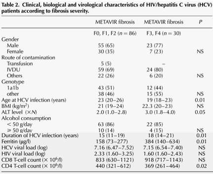
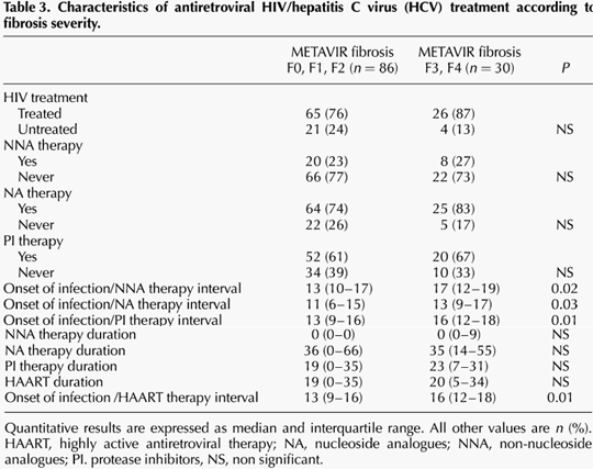
Among the 116 co-infected patients, 91 (78%) were receiving HAART at the time of liver biopsy. Immunological, virological and treatment data are shown in Tables 2 and 3. No difference was observed between the two groups regarding alcohol consumption, HCV genotype, HIV and HCV viral load, proportion of patients receiving antiretroviral treatment. Patients in group 2 had higher transaminase and ferritin levels and a lower CD4 T-cell count. No association was found between the Metavir score and exposure to didanosine, indinavir, ritonavir and nevirapine. However a trend towards a higher score appeared for stavudine (P = 0.058). Median duration of HAART as well as median duration of treatment with nucleoside analogues (NA), non-nucleoside analogues (NNA) and protease inhibitors (PI) were comparable in both groups. The delay between the presumed contamination and HAART initiation was significantly longer for patients from group 2 (16 versus 13 years; P = 0.01). Similar results were obtained when considering NA (13 versus 11 years; P = 0.03), NNA (17 versus 13 years; P = 0.02) and PI (16 versus 13 years; P = 0.01). However, after adjusting in a logistic regression model for duration of disease, age at contamination and CD4 T-cell count, none of these variables remained statistically significant.
|
|
| |
| |
 |
|
| |
| |
|
|
| |
| |
 |
|
| |
| |
A second analysis was performed excluding patients with a METAVIR score of F4 (n = 19), in order to avoid a possible measuring bias linked to the uncertain date of appearance of liver cirrhosis. The results of this analysis showed the same differences between the two groups regarding age at contamination, transaminase and ferritin levels. Median duration of HAART was higher in group 1 than in group 2 (19 versus 8 months, P = 0.075). Median interval between the presumed contamination and HAART initiation was higher in group 2 (17 versus 13 years; P = 0.04), as well as the median interval between the presumed contamination and NA (14 versus 11 years; P = 0.04), NNA (17 versus 13 years; P = 0.07) and PI initiation (17 versus 13 years; P = 0.04). We performed several logistic stepwise regression model analyses with duration of disease, age at contamination and CD4 T-cell count variables entered in the model at the initial step. Results show that patients in group 1 with moderate fibrosis had a significantly shorter delay from presumed date of contamination until HAART treatment initiation compared with those in group 2 (adjOR = 1.16; 95% CI, 1.01-1.32; P = 0.03) and longer duration of HAART (adjOR = 0.96; 95% CI, 0.91-1.0; P = 0.06). Among the different classes of antiretroviral regimens, only the median interval between presumed date of contamination and PI initiation remained associated with moderate fibrosis (adjOR = 1.15; 95% CI, 1.01-1.31; P = 0.03).
We conducted a third analysis on non-cirrhotic patients focusing on the mean rate of fibrosis progression according to the various characteristics of the co-infected patients. In a multiple regression model adjusting for duration of disease, age at contamination and CD4 T-cell count, mean rate of fibrosis progression was significantly slower among patients for whom duration of HAART was longer (β = -0.190; P = 0.02) and for whom the delay from presumed time of contamination until HAART initiation was shorter (β = 0.694; P = 0.02).
|
|
| |
| |
|
|
|