| |
HCV Did Not Persist in Patients with SVR
|
| |
| |
"Long-term follow-up after successful interferon therapy of acute hepatitis C"
Hepatology
June 2004
Virological Outcome
All patients included in this study had been HCV RNA-negative at the end of IFN--2b therapy and after 24 weeks of follow-up.
Thereafter, all 31 individuals were serum HCV RNA-negative by COBAS Amplicor (Roche Diagnostics, Mannheim, Germany) assay (detection limit 600 IU/mL) for the entire follow-up and at their most recent visit (range, 52-224 weeks).
For 20 patients, serum was available to investigate low levels of HCV RNA with the more sensitive TMA assay (detection limit 5-10 IU/mL). All samples tested were HCV RNA-negative. Finally, RNA could be isolated from PBMC of 15 patients. HCV RNA could not be detected in the PBMC of any of these individuals (Table 2).
AUTHOR CONCLUSION:
The most important finding of this study was that none of the patients had evidence of a late virological relapse. All patients remained serum HCV RNA-negative as determined by standard HCV PCR. Also important, there was no evidence for low levels of persisting virus using the more sensitive TMA assay. In addition, all patients tested were HCV-RNA-negative in PBMC. However, we cannot exclude persistence of low levels of HCV in the liver because there was no indication to perform liver biopsies in patients who were serum HCV -RNA-negative for 2 to 4 years.
Authors: Johannes Wiegand 1, Elmar Jäckel 1, Markus Cornberg 1, Holger Hinrichsen 2, Manfred Dietrich 3, Julian Kroeger 4, Wolfgang P. Fritsch 5, Anne Kubitschke 1, Nuray Aslan 1, Hans L. Tillmann 1, Michael Peter Manns 1, Heiner Wedemeyer 1 *
1Department of Gastroenterology, Hepatology and Endocrinology, Hannover Medical School, Hannover, Germany
2Medizinische Klinik, Universität Kiel (Health Clinic, University of Kiel), Kiel, Germany
3Bernhard-Nocht Institut Hamburg (Bernhard-Nocht Institute Hamburg), Hamburg, Germany
4Medzinische Klinik II, Universität des Saarlandes (University of Saarlandes), Hamburg/Saar, Germany
5Städtisches Krankenhaus Hildesheim (State Hospital), Hildesheim, Germany
ABSTRACT
Early treatment of acute hepatitis C infection with interferon alfa-2b (IFN--2b) prevents chronicity in almost all patients. So far, no data are available on the long-term outcome after interferon (IFN) therapy of acute hepatitis C.
The aim of this study was to assess the clinical, virological, and immunological long-term outcome of 31 successfully treated patients (65% genotype 1, 13% genotype 2/3, 23% unknown genotype) with acute hepatitis C infection who were followed for a median of 135 weeks (52-224 weeks) after end of therapy. None of the individuals had clinical evidence of liver disease.
Alanine aminotransferase (ALT) levels were normal in all but 1 patient. Serum hepatitis C virus (HCV) RNA was negative throughout follow-up, even when investigated with the highly sensitive transcription-mediated amplification (TMA) assay (cutoff 5-10 IU/mL).
In addition, no HCV RNA was detected in peripheral blood mononuclear cells (PBMC) of 15 cases tested.
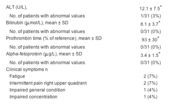
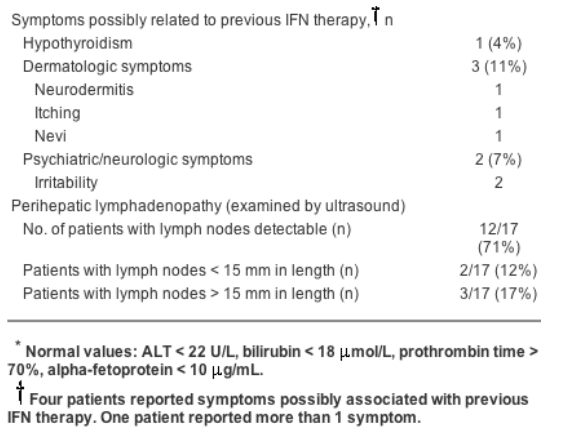
Table 2. Clinical and Virological Outcome
HCV RNA (PCR)-negative in serum (Cobas Amplicor-detection limit 600 IU/mL) 31/31 (100%)
HCV RNA (PCR) negative in serum (VERSANT/TMA assays, detection limit 5-10 IU/mL) 20/20 (100%)
HCV RNA-negative in PBMC 15/15 (100%)

The patients' overall quality-of-life scores as determined by the SF-36 questionnaire did not differ from the German reference control cohort. Ex vivo interferon gamma (IFN-) ELISPOT analysis detected HCV-specific CD4+ T-helper cell reactivity in only 35% of cases, whereas HCV-specific CD8+ T-cell responses were found in 4 of 5 HLA-A2-positive individuals. Anti-HCV antibody levels decreased significantly during and after therapy in all individuals.
In conclusion, early treatment of symptomatic acute hepatitis C with IFN--2b leads to a long-term virological, biochemical, and clinical response. Waning of anti-HCV humoral immunity and presence of HCV-specific CD8+ (but not CD4+) T cells highlights the complexity of T-cell and B-cell memory to HCV, which might be significantly altered by IFN treatment.
AUTHOR DISCUSSION
Early treatment of symptomatic acute hepatitis C infection with IFN--2b for 24 weeks leads to a virological response in 98% of cases.[5] The aim of this study was to assess the clinical, virological, and immunological outcome of patients after a median follow-up period of 2.6 years after the end of IFN-a-2b monotherapy.
The most important finding of this study was that none of the patients had evidence of a late virological relapse. All patients remained serum HCV RNA-negative as determined by standard HCV PCR. Also important, there was no evidence for low levels of persisting virus using the more sensitive TMA assay. In addition, all patients tested were HCV-RNA-negative in PBMC. However, we cannot exclude persistence of low levels of HCV in the liver because there was no indication to perform liver biopsies in patients who were serum HCV -RNA-negative for 2 to 4 years. The virological response was accompanied by a biochemical response. ALT values were normal in all but 1 patient. This patients minimal increased ALT might reflect steatohepatitis in the presence of obesity (BMI 28 kg/m2); there was no evidence of diabetes mellitus in this case.
Analysis of the SF-36 questionnaire revealed that the study populations quality of life did not differ from that of the German reference population in all 8 categories. However, it is not possible to judge whether quality-of-life status changed after the successful IFN therapy because there was no evaluation before, during, or right after treatment. It is also not known whether the patients will develop a more impaired health condition in further follow-up. Patients who had spontaneously cleared HCV from serum and who were followed for 15 years still demonstrated impaired quality-of-life scores, thereby supporting potential long-term effects of HCV on mental health status.[24]
Although the clinical and virological long-term outcome after IFN-a-2b monotherapy seems to be excellent, it is important to note that there were possible IFN-related long-term side effects in 14% of the patients. About 15% to 30% of patients with acute hepatitis C show a self-limited course of the disease.[2][25] These patients may be unnecessarily at risk to develop IFN-related long-term side effects when being treated. However, there are no sufficient predictive parameters available yet to identify patients with self-limited acute hepatitis C prior to treatment. The kinetics of HCV RNA decline during the first weeks of symptomatic infection may indicate self-limited disease,[26] but it must be pointed out that the mean time from exposure to HCV RNA negativity was 77 days in the study of Hofer et al.[26] whereas the average time from infection until the start of therapy was 89 days in our previous trial.[5] Thus, Hofer's patients would not have been included in our study. However, our data imply that patients should be informed extensively about potential risks associated with IFN- therapy before treatment is initiated. An alternative approach to managing symptomatic acute hepatitis C infection is to treat only those patients who are HCV-RNA-positive 12 weeks after the onset of symptoms to avoid unnecessary therapy. This concept may also lead to sustained virological response rates of up to 90%.[27] However, there are no data available concerning the long-term results of the wait-and-see strategy. A more comprehensive overview on different strategies for the management of acute hepatitis C has been recently published.[28]
We also analyzed the HCV-specific humoral and cellular immune response after long-term recovery from acute hepatitis C infection. Interestingly, after an early increase of the OD 405 of the anti-HCV-ELISA, we observed a biphasic decline of the HCV-specific humoral immune response. The rapid decrease of the anti-HCV antibodies until the end of the IFN therapy could be caused by the fast clearance of HCV-RNA, leading to an absence of viral antigen required for stimulation of B-cells. Alternatively, IFN-a could also have inhibited B-cell proliferation. After the end of therapy, OD 405 levels further declined, potentially leading to an earlier disappearance of anti-HCV antibodies compared to previous reports on patients who had recovered spontaneously from acute HCV infection without antiviral treatment.[11]
Kinetics of humoral responses during antiviral therapy did differ between patients with acute HCV infection and patients being treated for chronic hepatitis C. First, pretreatment antibody levels were higher in chronic infection; this is in line with previous reports.[29] Second, the early OD increase during treatment was more pronounced in patients with acute HCV, although pretreatment viral load and HCV-RNA decline during therapy were similar in both groups. Thus, the OD rise may be explained not only by a simple titration phenomenon due to drop in antigen load but also by differences in responsiveness of B-cells to type I IFN stimulation. Third, during therapy, antibody levels did not decline in patients being treated successfully for chronic hepatitis C but remained at high levels even after the end of therapy. The reasons for these observations are not clarified yet and remain to be investigated.
The cellular immune response has been shown to play a crucial role in the outcome of acute HCV infection. Strong CTL and CD4+ T-cell responses are associated with spontaneous clearance of the virus, while T-cell responses are very weak or even absent in chronic hepatitis C.[30-39] A maintained CD4+ T-cell response is a prerequisite for prolonged HCV-RNA negativity and protection from secondary infection,[40] while a loss of CD4+ T-cell reactivity in the acute phase of infection is accompanied with a reoccurrence of HCV RNA after initial viral clearance.[27] Using assay techniques identical to those applied in the present study, we usually detect HCV-specific CD4+ and CD8+ T cells in less than 10% of chronic HCV patients (data not shown). However, in IFN-recovered patients, responses were now detected only slightly more often, because HCV-specific CD4+ T cells were absent in about two-thirds of patients at their most recent follow-up. These data imply that there may be no longlasting CD4+ T-cell immune response against HCV after IFN-induced clearance of acute hepatitis C. The results are supported by our findings of weak CD4+ T-cell responses in the control patients recovered from chronic hepatitis C. Although some studies have suggested a restoration of T-cell responses in chronic hepatitis C by successful antiviral therapy,[41][42] no data are available for the investigation of HCV-specific cellular immunity later than 24 weeks after the end of therapy.
The lack of low levels of persisting viral RNA could be an explanation for the observed vanishing of CD4+ T-cell responses because it has been reported that CD4+ T-cell activity depends on restimulation with low levels of persisting antigen.[43] The degree of CD4+ memory T-cell persistence also depends on the initial burst size of immune responses during early infection[44]; however, this is not known for our patients. CD4+ T-cell responses observed in our study were multispecific and showed a broader spectrum than those in earlier studies of patients with self-limited acute hepatitis C.[45]
In contrast to the CD4+ T-cell immune response, an HCV-specific CD8+ T-cell immunity could be observed in 4 of 5 HLA-A2-positive patients. HCV-specific CD8+ T-cell memory has recently been proven to be essential for protection against persistent HCV infection on reexposure to HCV.[46] The positive CD8+ T-cell response in the present study does not necessarily seem to be dependent on CD4+ help,[47] although more patients are needed to draw final conclusions. However, the presence of CD8+ responses in our control group was also not accompanied by strong HCV-specific CD4+ responses. The rapid decline of the viral load at the beginning of therapy may have prevented an initial dysfunction and stunning of CD8+ T -cells; this can be observed in the initial phase of acute hepatitis C with high levels of antigen.[37] The level of CD8+ T cells correlates with viral clearance and outcome of acute hepatitis C infection, but the high level of CD8+ T-cell immunity is not preserved after the first months in untreated patients with acute hepatitis C.[37][38] Interestingly, patients without CD8+ T-cell response in the first 6 months after the onset of disease start to produce a specific immunity in the following months after the initiation of specific antiviral therapy.[39] Overall, these data support the hypothesis that IFN-a stimulates the induction of virus-specific CD8+ T cells.
In summary, the data from the present study prove the long-term efficacy of early IFN-a-2b therapy in patients with acute hepatitis C. However, the excellent treatment success may be accompanied by IFN-related side effects in some patients. In addition, this study gives evidence that IFN-a treatment may significantly influence the evolution of both cellular and humoral memory immune responses; this could have consequences if reexposure to HCV occurs.
Results
Clinical Outcome
Thirty-one patients with a history of acute hepatitis C were studied. All of them were HCV RNA-negative 24 weeks after cessation of therapy (sustained response). Patients were followed thereafter for a median of 135 weeks (range, 52-224 weeks) after the end of therapy.
None of the patients had clinical evidence of liver disease at their most recent visit. Complete biochemical response (aminotransferases within normal limits) was achieved in all but 1 patient, who had only minimally increased alanine aminotransferase (ALT) levels (25 U/L: upper limit of normal 22 U/L). This patient had a body mass index (BMI) of 28 kg/m2 and showed signs of steatosis on ultrasound examination. Liver function as determined by bilirubin, albumin levels, and prothrombin time was normal in all individuals (Table 2).


At their most recent follow-up visit, 18 of 28 patients (64%) reported being completely healthy without any evidence of symptoms potentially associated with hepatitis or IFN therapy. Two patients presented with mild intermittent abdominal pain in the right upper quadrant; one of these patients had cholecystolithiasis as documented by ultrasound. Another 3 patients suffered from fatigue, associated with an impaired ability to concentrate in 2 cases. The third patient additionally had thalassemia. Four of the patients (14%) reported persistence of symptoms that might be related to the previous IFN therapy: thyroid dysfunction, n = 1; dermatologic symptoms, n = 3; neurological/psychiatric symptoms, n = 2. Details are summarized in Table 2.
Seventeen patients were examined by high-resolution abdominal ultrasound. None of them displayed sonographic signs of chronic liver disease. Portal-vein flow and the resistance index of the hepatic artery were normal in all individuals. Patients were also screened for lymph nodes in the hepatoduodenal ligament because in the absence of liver biopsies, enlarged lymph nodes in the hepatoduodenal ligament have been shown to reflect intrahepatic inflammatory processes.[22][23] Importantly, lymph nodes larger than 15 mm were detected in only 3 patients (18%). In addition, there was no serological or clinical evidence of autoimmune liver disease; all patients tested negative for ANA, AMA, SMA, LKM, and SLA autoantibodies.
The quality-of-life analysis was assessed using the SF-36 questionnaire. The study population showed similar scores compared to those of the general German control group. The results were within the standard deviation of the controls in each of 8 categories.
Immunological Outcome
Anti-HCV-Specific Humoral Immune Responses
To analyze the strength of the HCV-specific humoral immune response, the optical density (OD 405) of anti-HCV enzyme-linked immunosorbent assay (ELISA) (AxSYM HCV version 3.0; Abbott Laboratories) was investigated over time. In all individuals there was a significant increase of the OD 405 until week 4 of therapy (Fig. 2). Thereafter, the OD values declined in a biphasic pattern. The decrease was more pronounced until the end of therapy; thereafter, it declined more slowly to a level below the initial value obtained before treatment (Fig. 2). In some cases, the OD 405 was close to the detection limit of the assay at the most recent follow-up visit. The initial increase of the OD 405 during the first 4 weeks of treatment of acute hepatitis C was always accompanied by a decrease of serum HCV RNA (Fig. 3). In contrast, kinetics of anti-HCV OD 405 values showed different patterns during successful IFN plus ribavirin combination therapy of chronic hepatitis C. While there was only a minor initial increase during the early phase of treatment of chronic HCV infection, OD levels did not decline thereafter and remained above the levels obtained before therapy.
The specificity of the humoral immune response was multispecific and targeted against both structural and nonstructural proteins prior to therapy. The recognition of core proteins decreased during the time course of follow-up, but the recognition of NS3 seemed to be more preserved (Fig. 4). The absolute frequency of recognition was higher for NS3 than for the core proteins. Compared to these proteins, the envelope protein E2 and NS5 were rarely recognized at the different time points.
MHC Class II-Restricted HCV-Specific T-cell Responses
To analyze MHC class II-restricted CD4+ T-cell responses to HCV proteins, PBMC were tested for IFN- production using the ELISPOT technique. Thirteen patients (65%) did not show IFN- responses at their latest follow-up visit. Six patients (30%) recognized 1 or 2 of the antigens tested; only 1 patient (5%) showed reaction against more than 2 antigens (Fig. 5A). The IFN- responses of the 7 responding patients were multispecific: no protein was recognized significantly more frequently than others (Fig. 5B). The frequency of specific spots per antigen ranged between 15 and 75 IFN--producing cells per 1 × 10⁶ PBMC. There was no correlation between frequency or strength of cellular immune responses as determined by the absolute number of HCV-specific T cells and time of follow-up after end of therapy.
Similar frequencies of HCV-specific CD4+ T-cell responses were observed in control individuals who had cleared chronic HCV infection. There was also no significant difference regarding the specificity of responses (Figs. 5C and D) or the absolute number of specific spots obtained in the ELISPOT assay.
MHC Class I-Restricted HCV-Specific T-cell Responses
PBMC for the analysis of MHC class I-restricted immune responses were available in 16 individuals. Five patients (31%) were HLA-A2-positive. HLA-A2-negative individuals did not show significant responses in the ELISPOT assay against the 7 tested HLA-A2-restricted peptides. In contrast, In 4 of 5 HLA-A2-positive patients (80%), in contrast, IFN- production could be observed after stimulation of PBMC with HLA-A2-restricted HCV-specific peptides (Fig. 6A). One patient recognized 4 of 7 different peptides tested; the other 3 patients recognized 1 or 2 peptides. Peptides E1-257 and core-178 were recognized most frequently; stimulation with peptides HCV core-35 and HCV core-131 did not cause significant IFN- production of CD8+ T cells (Fig. 6B). Of patients with detectable HCV-specific CD8+ T-cell responses, 3 of 4 also mounted significant CD4+ T-cell reaction against the tested proteins (data not shown). The median time of follow-up of the five HLA-A2-positive patients was 180 weeks. In control patients recovered from chronic HCV infection after IFN-based therapies, 4 of 9 HLA-A2-positive individuals mounted responses to 1 to 3 of the epitopes tested (Figs. 6C and D). There was no correlation between HCV-specific CD4+ and CD8+ T-cell responses in this group. If positive results were obtained, there was no difference in the absolute frequency of antigen-specific CD8+ T cells between patients recovered from acute HCV and those recovered from chronic HCV; frequency ranged between 15 and 45 IFN--producing cells per 1 × 10⁶ PBMC.
Article Text
Chronic hepatitis C virus (HCV) infection is one of the leading causes of chronic liver disease, affecting about 170 million people worldwide.[1] Acute infection with HCV leads to a chronic course in 55% to 85% of cases.[2] Although chronic HCV infection can be cured in 54% to 56% of cases with pegylated interferon alfa (IFN-) and ribavirin,[3][4] there is still no successful treatment option available for the majority of patients infected with HCV genotype 1. Thus, prevention of HCV chronicity seems to be desirable. Early treatment of acute symptomatic HCV infection with interferon alfa-2b (IFN--2b) monotherapy has been shown to be very successful: 98% of cases achieved a sustained virological response after a treatment period of 24 weeks.[5] So far, there are no data available concerning the long-term outcome after treatment of acute hepatitis C with IFN-. This might be of special interest because late relapses more than 6 months after spontaneous clearance of acute HCV infection have been described.[6] Therefore, the aim of this study was to assess the clinical, virological, and immunological outcome of 31 patients who were followed for 52 to 224 weeks after end of monotherapy with IFN--2b.
We also investigated HCV-specific humoral and cellular immune responses in these patients. Immunological memory to HCV has been discussed controversially in recent years.[7-10] While anti-HCV antibodies are thought to decline after clearance,[11] there are conflicting data on the strength and specificity of HCV-specific CD4+ and CD8+ T-cell responses.[11-14] In addition, it is not known whether antiviral therapy with IFN- might influence the evolution of virus-specific cellular immune responses. However, detailed knowledge of T-cell memory might be of importance for the development of vaccines against HCV.
Patients and Methods
Thirty-one patients were included in this analysis. All patients had a history of acute HCV infection and were successfully treated with IFN- monotherapy. Twenty-four patients were treated within the study reported by Jaeckel et al.[5] In addition, 7 patients were included who were treated with the same regimen but outside the trial. Treatment success was defined as sustained virological response (HCV-RNA [polymerase chain reaction]-negative 24 weeks after end of therapy, measured by COBAS Amplicor, version 2.0; Roche Diagnostics, Mannheim, Germany; detection limit 600 copies HCV RNA/mL). The exact definition of acute hepatitis C and criteria for indication for therapy have been described in detail.[5]
Eighteen patients were examined at Hannover Medical School; 13 patients were seen by their local physician and asked for their history using a questionnaire created for this study. This questionnaire covered the following topics: current health status, change of health status since IFN therapy, alcohol consumption, smoking habits or illicit drug use, concomitant diseases, body weight and height, current medication, and design and duration of IFN therapy. In Hannover, abdominal ultrasound examination and evaluation of autoantibodies (ANA, AMA, SMA, LKM, and SLA) were offered to the patients. Serum levels of HCV RNA were centrally tested by quantitative polymerase chain reaction (PCR) using COBAS Amplicor, version 2.0 (Roche Diagnostics; detection limit 600 IU HCV RNA/mL) at Hannover Medical School. Biochemical and hematological tests were performed locally.
The control group for T-cell analysis consisted of 13 individuals who had cleared chronic HCV infection after interferon (IFN)-based therapies. Patients were studied after a median of 56 weeks after the end of therapy (range, 24-262 weeks). All control patients were treated at Hannover Medical School and were constantly HCV RNA-negative in all regular follow-up visits. Control patients had no evidence of liver disease or other concomitant illnesses.
Optical density (OD) of anti-HCV antibodies was centrally analyzed with microparticle enzyme immunoassay AxSYM HCV version 3.0 (Abbott Laboratories, Abbott Park, IL) at weeks 0, 2, 4, 12, 24, and 48 and at the most recent time point after initiation of therapy. Antibody reactivity against HCV-specific antigens was evaluated using the immunoblot assay INNO-LIA HCV III Update (Innogenetics, Ghent, Belgium) at weeks 0 and 48 and at the most recent time point after the initiation of therapy. Baseline characteristics are summarized in Table 1.
Table 1. Baseline Characteristics of the Study Cohort
Patient Characteristics (N = 31)
Sex, n (%)
Male 15 (48%)
Female 16 (52%)
Median age, y (range) 40.0 (25-73)
Median body mass index kg/m2 (range) 24.8 (18.9-32.8)
HCV genotype prior to therapy, n (%)
Genotype 1 20 (65%)
Genotype 2/3 4 (13%)
Unknown 7 (23%)
Mode of infection, n (%)
Sexual 3 (10%)
Medical procedure 6 (20%)
Drug abuse 5 (16%)
Needle stick injury 9 (29%)
Unknown 8 (26%)
Median time of follow-up after end of therapy, wk (range) 135 (62-224)
Isolation of PBMC
Peripheral blood mononuclear cells (PBMC) were isolated as previously described.[14] Blood samples from patients not examined at Hannover Medical School were shipped at room temperature, and lymphocyte isolation was performed a maximum of 2 days after bleeding. Cell viability and functional fitness of shipped samples were ensured by staining with trypan blue and T-cell assays against control antigens. Lymphocytes were stored using cryopreservation in freezing medium (25 mL RPMI 1640 with 2 mmol L-glutamine, 50 mL fetal calf serum, and 8.3 mL DMSO) until further use.
Enzyme-Linked Immunospot (ELISPOT) Assay for CD4+ T-cell Immune Response
Cryopreserved PBMC (2 × 10⁵ up to 8 × 10⁵) were thawed and incubated overnight in duplicate cultures of 100 L RPMI 1640, 51 AB serum, and 2 mmol L-glutamine, together with HCV proteins (core, NS3, NS4, NS5, helicase; Microgen, Munich, Germany) 1 ug/mL in a 96-well plate (Microgen) as previously described.[15] In brief, a second 96-well plate (Millititer; Millipore, Bedford, MA) was coated with 100 uL (0.5 g/mL) anti-human interferon gamma (IFN-; Endogen, Woburn, MA) at 4°C overnight and washed 4 times with 200 uL phosphate-buffered solution The plate was blocked with 100 uL RPMI, 1% Bovine saline albumin (Sigma, St. Louis, MO) for 1 hour at 25°C and washed again with PBS. The preincubated cells were transferred to the anti-human IFN--coated plate and incubated overnight. These plates were washed 7 times and incubated overnight with 100 uL (1 ug/mL) biotin-conjugated anti-human -IFN-y (Endogen). After 4 washes, streptavidin-alkaline phosphatase (AP) (1:2000 in PBS; BioRad, Hercules, CA) was added for 2 hours. Finally, the plate was washed again 4 times with PBS and developed with freshly prepared NBT/BICPsolution (BioRad). The reaction was stopped with distilled water. The spots were counted using an automated ELISPOT reader (A-EL-VIS Elispot Reader; Hannover, Germany). The number of spots in the absence of antigen was less than 5 in 18 patients, between 5 and 10 in 2 patients and more than 10 in 0 patients. The number of specific spots was determined by subtracting the number of spots in the absence of antigen from the number of spots in the presence of antigen. Responses were considered to be positive if more than 5 specific spots were detected and if the number of spots in the presence of antigen was at least 2-fold greater than the number of spots in the absence of antigen. Positive controls consisted of phytohemagglutinin (1 ug/mL; Murex Biotech Limited, Dartford, England) and tetanus toxoid (50 ug/mL; Calbiochem, Merck Biosciences, Schwalbach, Germany).
ELISPOT Assay for CD8+ T-cell Immune Response
Seven previously described HLA-A2-restricted HCV peptides (core-35 YLLPRRGPRL, core-132 DLMGYIPLV, core-178 LLALLSCLTV, E1-257 QLRRHIDLLV, NS3-1073 CVNGVCWTV, NS3-1169 LLCPAGHAV, and NS3-1406 KLVALGINAV)[16] and the HLA-A2-restricted influenza A virus matrix peptide GILGFVFTL[17] were selected. Peptides were synthesized and HPLC purified to more than 90% purity at Research Genetics, Huntsville, AL.
Peptides were used in a final concentration of 10 g/mL. Cryopreserved PBMC (2 × 10⁵ to 5 × 10⁵) were incubated overnight in duplicate cultures of 100 L RPMI 1640, 5% AB, and 2 mmol L-glutamine in a 96-well plate (Nunc, Roskilde, Denmark). PBMC were transferred to an IFN- ELISPOT plate and incubated with peptides and 5 × 10⁴ HLA-A2-positive T2 cells overnight as described in Patients and Methods for CD4+ T-cell responses. All the following procedures were identical to the description in Patients and Methods.
RNA Isolation From PBMC
HCV-RNA was isolated from PBMC with a minipreparation-kit (High Pure RNA Isolation Kit; Roche Diagnostics) according to manufacturer's protocol. The HCV RNA reverse-transcriptase PCR was performed as described previously.[18]
Transcription-Mediated Amplification (MA) Assay
The TMA assay (detection limit 5-10 IU/mL)[19] was performed at the Christian-Albrechts-Universität Kiel, Germany using the VERSANT HCV-RNA Qualitative Assay (Bayer, Tarrytown, NY).
HLA Typing
HLA typing was done by PCR as previously described.[20]
Assessment of Quality of Life
The Medical Outcomes Study (MOS) 36-item short-form health survey (SF-36)[21] was used and answered by 27 patients.
Statistical Analysis
Students t tests were used, and differences with a P value less than .05 were considered statistically significant.
Ethics
This study was part of an approved protocol reviewed by the Ethics Committee of Hannover Medical School. Informed consent was given by each patient.
|
|
| |
| |
|
|
|