 |
|
|
| |
Heart Disease/Inflammation/Activation - T-cell Activation and E-Selectin Associated with Coronary Plaque in HIV-infected Youth
|
| |
| |
Reported by Jules Levin
Oct 6-8, 2014
16th International Workshop on Co-Morbidities and Adverse Drug Reactions in HIV
AB Unsal, KZ Abd-Elmoniem, A Rupert, J Kovacs, JB Purdy, R Hazra, AM Gharib, CM Hadigan
National Institutes of Health
Bethesda, MD
"Conclusions: Prior investigation in HIV-infected adults has identified an association between increased carotid lesions and T-cell activation markers.
- Individuals infected with HIV early in life have higher levels of immune activation, inflammation, and circulating adhesion molecules than controls
- Viral suppression of HIV helps reduce but not completely reverse the chronic inflammatory state of HIV
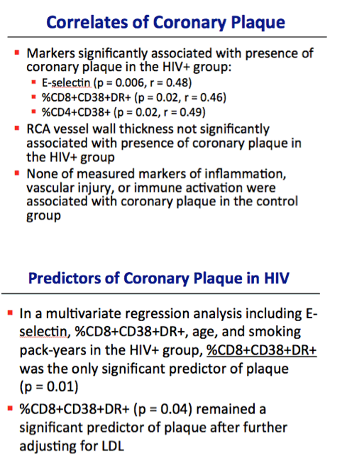
we identified a significant relationship between increased coronary plaque and levels of activated T-cells in young adults with life-long HIV. Further, soluble E-selectin, which has also been linked with carotid artery plaque and atherosclerosis was positively correlated with coronary plaque in the present study. The presence of increased circulating adhesion molecules and markers of immune activation may be early predictors of atherosclerosis in young adults infected with HIV in early life."
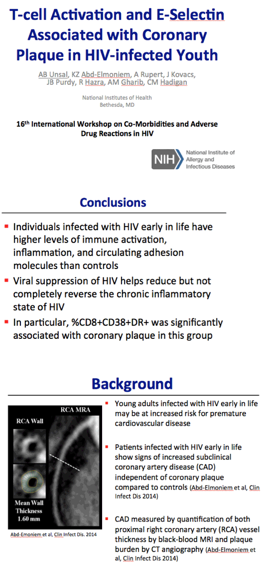
Coronary vessel wall thickness by BB-MRI -> image in an HIV-infected patient is shown to the left. Novel MRI technique that allows measurement of the actual wall thickness by silencing the lumen signal and keeping the MRI signal from the wall. May be more indicative of CVD events than cIMT or lumen area.
Program abstract:
T-cell activation and E-selectin associated with coronary plaque in HIV-infected youth
AB Unsal1, KZ Abd-Elmoniem1, A Rupert1, J Kovacs1, JB Purdy1, R Hazra1, AM Gharib1, CM Hadigan1
1National Institutes of Health, Bethesda, MD, USA
Aim: Adolescents and young adults infected with HIV early in life may be at increased risk of atherosclerotic cardiovascular disease. Recent research strongly suggests the importance of chronic inflammation and circulating adhesion molecules in the early pathogenesis of atherosclerosis, however, much has yet to be elucidated regarding the mechanism of vascular injury in this population. The aim of this study, therefore, was to measure biomarkers of cardiovascular injury and immune activation in relationship to coronary plaque burden in patients infected with HIV early in life.
Methods: 35 youth and young adults who acquired HIV early in life and 11 uninfected healthy controls were examined in this prospective cross-sectional study. All subjects and controls were free of active cardiovascular disease at the time of evaluation. CT angiography was utilized for coronary plaque quantification. Number of atherosclerotic plaques, calcified and non-calcified, was determined in each of the 17 American Heart Association (AHA) coronary segments.
Results: We studied HIV-infected subjects (mean age 22; range 15-29 years; 54% male) and 11 HIV-uninfected control subjects (mean age 25; 22-29 years; 27% male). No calcified plaque was found in either study group. There was no significant difference in number of plaque lesions between groups (HIV+ median 0, range 0-4, control median 0, range 0-7, P=0.08). Level of activated CD8 T-cells in the periphery, as measured by %CD8+CD38+DR+, was associated with increased coronary plaque in the HIV+ group (P=0.02) as was %CD8+CD38+ (P=0.005). In the CD4 population %CD4+CD38+ was also associated with increased coronary plaque (P=0.02), but not %CD4+CD38+DR+ (P=0.16). Further, E-selectin was significantly associated with plaque in the HIV+ subjects (P=0.006). There was no association between coronary plaque and levels of CRP, d-dimer, or homocysteine in either study population. Although P-selectin, sICAM-3, VCAM-1, TIMP-1 and MCP-1 levels were significantly elevated in HIV, these biomarkers were not related to plaque, nor were lipopolysaccharide binding protein levels. In a multivariate regression analysis of plaque adjusting for E-selectin, %CD8+CD38+DR+, age and smokingpack-years in the HIV group, %CD8+CD38+DR+ was the only significantly predictor of plaque (P=0.01). %CD8+CD38+DR+ remained a significant predictor of plaque (P=0.04) in the patients after further adjusting for LDL level.
Conclusions: Prior investigation in HIV-infected adults has identified an association between increased carotid lesions and T-cell activation markers. Here, we identified a significant relationship between increased coronary plaque and levels of activated T-cells in young adults with life-long HIV. Further, soluble E-selectin, which has also been linked with carotid artery plaque and atherosclerosis was positively correlated with coronary plaque in the present study. The presence of increased circulating adhesion molecules and markers of immune activation may be early predictors of atherosclerosis in young adults infected with HIV in early life.
1.Inflammation is integral in the formation of arterial plaque
1.Image on the right is a cross-sectional schematic showing arterial plaque formation from fatty streak formation through plaque rupture and thrombosis
2.What's really integral to this process is the role of inflammation - after initial injury to the endothelial cell lining, inflammation is the process that is both initiates and mediates the atherogenic process
2.On the left we see biomarkers associated with the plaque formation process - we have measurable markers that we can measure that may both represent different points in the process but also may be predictors of eventual plaque rupture and cardiovascular events. Since many of these biomarkers are associated with different stages of the atherogenic process, we may be able to understand where we are in the process by measuring biomarkers. Furthermore, these biomarkers may be predictive of future cardiovascular events.
3.Finally again on the right side shows the earliest onset of subclinical plaque formation - begins in the first decade of life. We can measure subclinical plaque formation in even younger populations of adults that would not be traditionally thought of as having plaque or CVD.
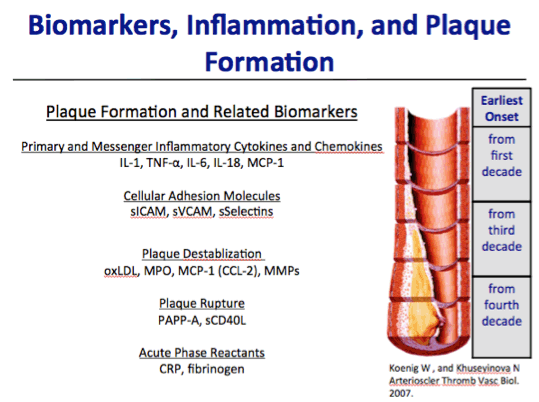
- Inflammation is linked w/ plaque formation
- pathophysiology of atherosclerosis: injury to endothelial cell (by smoking, HTN, idk) which causes inflammation, LDL in blood stream will lodge between basement membrane and eventually form foam cells - starts to collect and also causes more inflammation
- Inflammation causes more LDL to come until fibrin is finally deposition and we form a fibrous plaque (no more platelet aggregation); inflammation causes breakdown of the fibrous cap and eventually may rupture (forms a big clot, may cause embolization)
- Measuring biomarkers may provide us with information regarding the mechanism of CVD in our cohort and may also be predictors of future cardiovascular events
Pathophysiology of Atherosclerosis:
- Primarily involves the tunica intima in the beginning
- Have damage to the tunica intima via smoking, diabetes, HTN - but what else? Injury to one of the endothelial cells which releases vWF
- vWF connects platelets and then causes inflammation *** WE ARE ALREADY IN AN INFLAMMATORY STATE (causes vasodilation, increased blood flow, capillary leak, calls WBCs - macrophages**)
- LDL in bloodstream will then lodge between the basement membrane of the tunica intima and start to collect and then the LDL itself will cause more inflammation
- Fatty streak formation occurs w/ continued LDL deposition under the tunica intima, macrophages then become foam cells à continues as the inflammation gets worse, LDL continues to accumulate
- End process of inflammation - deposition of FIBRIN à development of a FIBROUS PLAQUE (will become an indolent process above the surface in the lumen of the bloodstream, no more platelet aggregation): all of the inflammation causes the deposition of fibrin so we end up with fibrous cap trying to enclose the big inflamed area but can have more LDL deposition and more inflammatory mediators
- With more inflammation, we end up getting degradation of the fibrous cap à thinner fibrous cap makes the plaque more prone to rupture and clinical events and the inflammatory "stew" all of a sudden spews out and activates more inflammatory factors, platelets, and the coagulation pathway will then form a big clot that can eventually embolize (causes ACS, unstable angina, CVA, peripheral vascular emboli - ischemic limb)
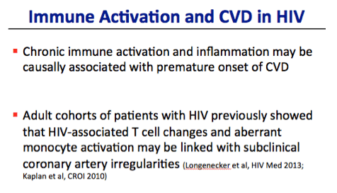
-Research in adults with HIV has shown that immune activation and inflammation associated with HIV may cause premature onset of CVD
-Research from the WIHS and SATURN cohorts show that
HIV-associated T-cell changes - as measured by immune activation and immune senescence - may be linked with subclinical abnormalities of the coronary artery
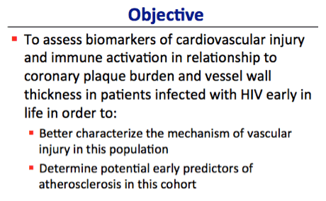
We were interested in studying the role of immune activation and biomarkers of vascular injury with coronary artery disease (measured by vessel wall thickness and plaque) in those patients with nearly lifetime HIV infection to: characterize vascular injury in this cohort and determine early predictors of CVD in this group
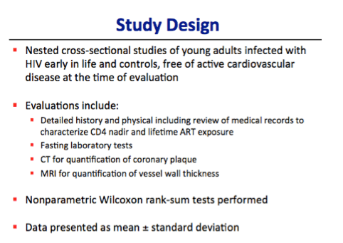
1.The study with this cohort of patients infected with HIV early in life is a 10-year longitudinal study with nested cross-sectional studies
2.We sex- and race-matched w/ controls all free of cardiovascular disease at the time of evaluation
1.Pt and controls are not age-matched due to radiation issues w/ CT - could not have controls under the age of 18
3.Evaluations at yearly visits include:
1.READ
4.Statistics - non-parametric Wilcoxon rank-sum tests were performed for univariate analyses and the data is presented as mean +/- standard deviation unless otherwise noted
Why is this MRI the best? - non-operator dependent, only technique to show the coronaries (non-invasive), not just showing the lumen but also the actual thickness of the vessel
BB-MRI - relies on nulling of the MR signal from the vascular lumen while retaining signal from the vascular wall, allowing better visualization of wall thickness
-Plaque was defined as any calcification involving the coronary artery or non-calcified soft tissue density abutting the coronary lumen causing irregularlity or significant luminal narrowing
-Evaluated 17 AHA segments for number of lesions, size, and amount of luminal narrowing
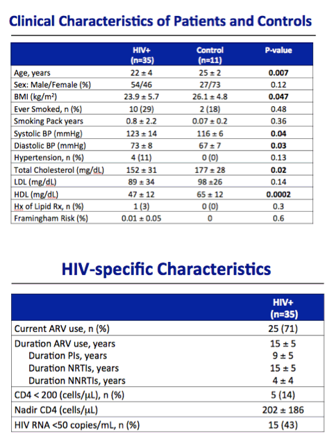
These characteristics are consistent with other young adult populations
Why is duration of ARV use diff than age? - many were infected very early on in the epidemic/pre-HAART era, limited access and some not diagnosed until years after their initial infection, have periods of non-adherence
Although these characteristics may not represent the adult HIV population, they are consistent with other cohorts of young adults with HIV
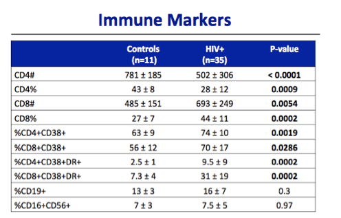
CD19+ = biomarker of B cell development
CD16+CD56 = markers on NK cells
Unsurprisingly, CD4 counts and percentages were significantly lower in the HIV population and CD8 counts and percentages were higher in the HIV population. Levels of activated T cells in the periphery - as shown by the 4 markers below - were also significantly higher in the HIV populations. CD19+, a marker of B cell development, and CD16+CD56+, a marker of NK cells, were not significantly different between the two groups.
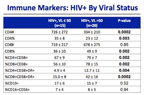
Upon splitting the HIV group into those that were viremic and not, we saw similar, unsurprising relationships - CD4 count/percent lower in viremic, levels of activated T cells in the periphery were significantly higher in the HIV group.
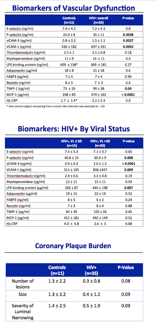
When we evaluated the coronary arteries for plaque, did not just look at number of plaque lesions but also looked at size and severity of luminal narrowing
1.For patients and controls, all of the plaque found was non-calcified (higher risk for plaque rupture)
Although not statistically different, there was more non-obstructive, non-calcified plaque in the controls than in the HIV+ population (most likely due to slightly higher age in the control group)
**** NO PLAQUE CAUSED LUMINAL NARROWING, NO PLAQUE WAS CALCIFIED (I.E. all were non-calcified which is more at risk for eventual plaque rupture)
|
| |
|
|
|
|
|