 |
|
|
| |
Magnetic Resonance Spectroscopy Evidence of Accelerated Aging in Virally-Suppressed HIV
|
| |
| |
Download the PDF here
Download the PDF here
Download the PDF here
10th International Workshop on HIV and Aging, October 10-11, 2019, New York
Reported by Jules Levin
Poster 29
Thao Tran1, Karen Chu1, Ke Wei1, Kimberly Shriner2, Kevin King1
1 Advanced Imaging & Spectroscopy Center, Huntington Medical Research Institutes, Pasadena, CA
2 Phil Simon Clinic, Huntington Memorial Hospital, Pasadena, CA
poser slides resume following some additional data I include below relevant to this topic.
--------------------------------

https://academic.oup.com/ofid/article/5/10/ofy243/5104905?searchresult=1
pdf attached above
Abstract
Background
Combination antiretroviral therapy (cART) has transformed HIV into a manageable but complex chronic disease, in which it is uncertain which brain insults may relate to age vs initial disease severity. We evaluate N-acetyl-aspartate/creatine (NAA/Cr), white matter hyperintensities (WMH), and mean cortical thickness to identify which subclinical markers of brain insult best relate to CD4 nadir and aging. This is a prospective study of the association between brain markers with age and initial infection severity, based on CD4 nadir, in chronic HIV patients.
Methods
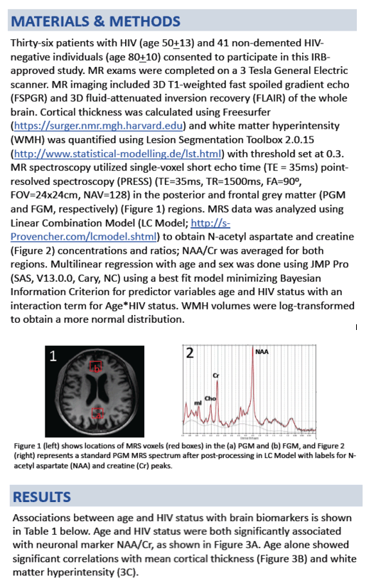
Thirty-seven chronic HIV patients (age 25-77 years) with successful viral suppression were scanned on a GE 3T magnetic resonance imaging scanner to obtain NAA/Cr (standardized and averaged over 5 brain regions), log-transformed WMH volume, and mean cortical thickness. The brain measures were fitted with both CD4 nadir and age to evaluate the significance of their relationship.
Results
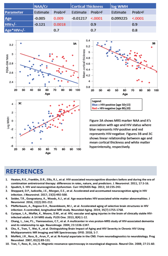
NAA/Cr, WMH, and cortical thickness were all correlated with age and CD4 nadir in unadjusted associations. Stepwise regression models showed that NAA/Cr alone best predicted CD4 nadir (β = 40.1 ± 13.3; P = .005), whereas WMH (β = 2.3 ± .9; P = .02) and mean cortical thickness (β = -2.7 ± 6.6; P < .0001) together produced the best model fit with age. NAA/Cr was higher for HIV stage 1 (CD4 nadir ≥ 500 cells/ μL; n = 15) compared with stage 2 (200 ≥ CD4 nadir < 500; n = 13) and stage 3 (CD4 nadir < 200; n = 9; P < .01 for both).
Conclusions
In patients with effectively suppressed HIV, NAA reflects the subclinical brain impact of initial disease severity related to development of even mild immune compromise, whereas cortical thickness and WMH volume are useful to evaluate age-related changes.
Regionally Specific Brain Volumetric and Cortical Thickness Changes in HIV-infected Patients in the HAART era
https://europepmc.org/articles/pmc5340610
pdf attached above
Abstract
Background
Cognitive impairment still occurs in a substantial subset of HIV-infected patients, despite effective viral suppression with highly active antiretroviral therapy (HAART). Structural brain changes may provide clues about the underlying pathophysiology. This study provides a detailed spatial characterization of the pattern and extent of brain volume changes associated with HIV, and relates these brain measures to cognitive ability and clinical variables.
Methods
Multiple novel neuroimaging techniques (deformation-based morphometry, voxel-based morphometry and cortical modeling) were used to assess regional brain volumes in 125 HIV-infected patients and 62 HIV-uninfected individuals. 90% of the HIV-infected patients were on stable HAART with a majority (75%) having plasma viral suppression. Brain volumetrics and cortical thickness estimates were compared between the HIV-infected and uninfected groups, and the relationships between these measures of brain volume and indices of current and past infection severity, central nervous system penetration of HAART, and cognitive performance were assessed.
Results
Regionally specific patterns of reduced thalamic and brainstem volumes, as well as reduced cortical thickness in the orbitofrontal cortex, cingulate gyrus, primary motor and sensory cortex, temporal, and frontal lobes were seen in HIV-infected patients compared to HIV-uninfected participants. Observed white matter loss and subcortical atrophy were associated with lower nadir CD4 cell counts, while reduction in cortical thickness was related to worse cognitive performance.
Conclusion
Our findings suggest that distinct mechanisms may underlie cortical and subcortical injury in people with HIV, and argues for the potential importance of early initiation of HAART to protect long term brain health.
The findings here support the hypothesis that subcortical structures are particularly vulnerable to the virus 33, and add spatial detail regarding the distribution of atrophy. In this study, we took advantage of multiple neuroimaging methods to investigate the spatial distribution of cortical and subcortical volume loss in a group of HIV-infected individuals in the HAART era. We observed significant reductions in cortical thickness throughout the cortex and WM tissue densities in subcortical structures, and worse cognitive performance in the HIV-infected cohort. HIV-infected patients with a history of more severe immunosuppression had significantly smaller subcortical volumes, but this variable was not related to cortical thickness reductions. In contrast, worse cognitive performance was associated with reduced regional cortical thickness but not subcortical volumes. The results presented provide a detailed characterization of the pattern and extent of brain injury in a diverse group of HIV-infected patients......The HIV-infected group performed significantly worse than demographically similar HIV-uninfected individuals on neuropsychological tests, adding to the growing body of evidence that suggests HIV-related cognitive impairment remains frequent despite effective treatment and minimal comorbidities 1. Regional cortical thickness reductions were related to poorer neuropsychological performance in the left lateral temporal pole, left inferior occipital, right lateral occipital and right inferior lateral frontal cortices, effects that presumably reflect the regions engaged by the particular set of cognitive tests administered. In contrast, subcortical volume changes were not significantly related to overall performance. These differential results may reflect the fact that the neuropsychological tests that were administered primarily assess cortical-hemispheric functions and might be less sensitive to subcortical dysfunction, a priori........To assess whether brain volume changes were influenced by variables related to the infection, brain volumes were correlated with nadir CD4, current CD4, viral load, and CPE measures within the HIV-infected group. VBM and DBM analysis revealed that HIV-infected patients with a history of more severe immunosuppression, indexed by nadir CD4, had significant reductions in WM density and smaller volumes in the brainstem, thalamus, caudate, putamen, globus pallidus, internal capsule and right frontal lobe, as well as greater enlargement of the third ventricle. This finding is consistent with the existing literature 9,11,37-40 supporting the theory that volume loss, in part, occurs during the time of untreated infection 8,12 suggesting that early initiation of HAART might be beneficial for long term brain integrity. Longitudinal data is required to determine whether these are static changes, arrested by HAART, or continue to develop despite HAART.
--------------
Longitudinal Trajectories of Brain Volume and Cortical Thickness in Treated and Untreated Primary Human Immunodeficiency Virus Infection
Clinical Infectious Diseases 1 December 2018
Pdf attached above
Abstract
Background
Human immunodeficiency virus (HIV) penetrates the brain in early infection. We used neuroimaging to longitudinally examine the impact of HIV and combination antiretroviral therapy (cART) on the brain in treated and untreated HIV-infected participants, starting in primary HIV infection (PHI).
Methods
Sixty-five participants, enrolled during PHI, underwent longitudinal magnetic resonance imaging, 30 of whom commenced cART during follow-up. Cross-sectional data from 16 patients with chronic HIV infection (CHI) and 19 HIV-uninfected participants were included for comparison. Brain volume and cortical thickness were estimated using tensor-based morphometry and cortical modeling, respectively. Mixed-effects models longitudinally mapped structural brain changes before and after cART. The relationship between brain morphometry estimates and blood and cerebrospinal fluid (CSF) biomarkers were also tested. Region-of-interest analyses were performed to compare brain morphometry estimates between the groups.
Results
Prior to cART, longer duration of untreated infection in PHI correlated with volume loss in the thalamus, caudate, and cerebellum, and with cortical thinning in the frontal and temporal lobes and cingulate cortex. After cART, no further volume loss was observed. However, small increases of cortical thickness in the frontal and temporal lobe correlated with longer cART duration. No correlations were observed with blood or CSF measures. The PHI group did not have different brain morphometric measures compared to the HIV-uninfected group, but had larger volumes in the thalamus, caudate, putamen, and cortical gray matter compared with CHI participants.
Conclusions
Subcortical atrophy and cortical thinning occur during untreated infection but may be arrested by cART. These findings emphasize the importance of early cART.
--------------------------------
|
| |
|
|
|
|
|