| |
Pioglitazone Improves Liver, Body Fat, Glucose
|
| |
| |
"A pilot study of pioglitazone treatment for nonalcoholic steatohepatitis" (fatty liver)
Improvements observed in body fat and glucose
Study found significant increases in total body fat: "...significant increases in total fat mass (+3.1 ± 3.6 kg, P =0.002), but little or no change in lean body mass (+0.2 ± 2.2 kg, P = not significant)..."
"...Mean body mass index increased 1.3 ± 1.7 kg/m2. Waist circumferences did not change, but waist-to-hip ratios decreased significantly. Whole body composition by DEXA showed that the weight gain was mostly due to an increase in adiposity, with significant increases in total fat mass (+3.1 ± 3.6 kg), but little or no change in lean body mass (+0.2 ± 2.2 kg, P = not significant). Weight gain was highly correlated with increase in total fat mass by DEXA ([r];=0.93). In contrast, liver fat and liver volume as assessed by MRI both decreased significantly..."
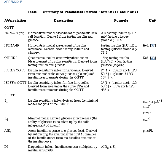
"...At baseline, 11 (61%) patients had impaired glucose tolerance, and two (11%) had values consistent with diabetes (2-hr glucose values 200 mg/dL). After treatment, only six subjects had impaired glucose tolerance, and no patient had 2-hr postglucose load values in the diabetic range. Insulin sensitivity indices as determined by homeostasis model assessment of insulin resistance (HOMA-IR) and quantitative insulin sensitivity check index (QUICKI) improved significantly after treatment....", but other measures of glucose did not improve (see text and table below).
"...Pioglitazone was generally well tolerated..."
"...No patient developed worsened serum ALT levels..."
"...No patient developed anemia or clinically apparent edema, reported side effects of pioglitazone...."
Hepatology
Volume 39, Issue 1, Pages 188-196
Kittichai Promrat 1, Glen Lutchman 1, Gabriel I. Uwaifo 2, Renee J. Freedman 2, Alejandro Soza 1, Theo Heller 1, Edward Doo 1, Marc Ghany 1, Ahalya Premkumar 4, Yoon Park 1, T. Jake Liang 1, Jack A. Yanovski 2, David E. Kleiner 3, Jay H. Hoofnagle 1 *§
1Liver Diseases Section, Digestive Diseases Branch, National Institute of Diabetes and Digestive and Kidney Diseases 2Unit on Growth and Obesity, Section on Women's Health, Developmental Endocrinology Branch, National Institute of Child Health and Development 3Laboratory of Pathology, National Cancer Institute
4Department of Diagnostic Radiology, Warren G. Magnuson Clinical Center, National Institutes of Health, DHHS, Bethesda, MD
ABSTRACT
Nonalcoholic steatohepatitis (NASH) is a common chronic liver disease for which there is no known effective therapy. A proportion of patients with NASH progress to advanced fibrosis and cirrhosis. NASH is considered one of the clinical features of the metabolic syndrome in which insulin resistance plays a central role.
This prospective study evaluates the role of insulin-sensitizing agent in treatment of NASH.
Eighteen nondiabetic patients with biopsy-proven NASH were treated with pioglitazone (30 mg daily) for 48 weeks. Tests of insulin sensitivity and body composition as well as liver biopsies were performed before and at the end of treatment.
By 48 weeks, serum alanine aminotransferase values fell to normal in 72% of patients. Hepatic fat content and size as determined by magnetic resonance imaging decreased, and glucose and free fatty acid sensitivity to insulin were uniformly improved. Histological features of steatosis, cellular injury, parenchymal inflammation, Mallory bodies, and fibrosis were significantly improved from baseline (all P < 0.05).
Using strict criteria, histological improvement occurred in two-thirds of patients. Pioglitazone was well tolerated; the main side effects were weight gain (averaging 4%) and an increase in total body adiposity. In conclusion, these results indicate that treatment with an insulin-sensitizing agent can lead to improvement in biochemical and histological features of NASH and support the role of insulin resistance in the pathogenesis of this disease. The long-term safety and benefits of
pioglitazone require further study.
Authors said--In this pilot study, we assessed whether pioglitazone would improve insulin sensitivity and affect biochemical and histological features of disease activity in nondiabetic patients with NASH. All 18 patients enrolled had improvements in serum ALT levels, which became normal in 72%. Liver histology showed improvements in the major features of NASH (steatosis, cell injury, inflammation, Mallory bodies, fibrosis), and two-thirds of the 18 patients reached the predetermined endpoint of histological improvement. The improvement in steatosis was also documented by hepatic imaging, in that MRI of the liver showed marked decreases in liver fat and liver volume. Most patients also had improvements in insulin sensitivity, as measured by oral and intravenous glucose tolerance tests, and surrogate serum markers including fasting insulin, C-peptide, and free fatty acid levels.
BACKGROUND
Nonalcoholic steatohepatitis (NASH) is a common but often silent chronic liver disease that resembles alcoholic liver disease clinically and histologically but occurs in persons who drink little or no alcohol.[1][2] NASH is part of a spectrum of nonalcoholic fatty liver disease that ranges from pure fatty liver (steatosis) to steatohepatitis and to cirrhosis.[3] The prevalence of NASH in the general population is unknown. Using elevated alanine aminotransferase (ALT) levels without an explainable cause as a surrogate for the diagnosis of nonalcoholic fatty liver disease, the prevalence of NASH in the United States was estimated to be 2.8%.[4] While once thought to be a benign condition, there is increasing evidence that NASH can lead to progressive fibrosis and eventually cirrhosis.[3][5-7] Indeed, a proportion of cases of cryptogenic cirrhosis may be burnt out NASH.[8] Furthermore, long-standing NASH with cirrhosis has been associated with the development of hepatocellular carcinoma.[9]
The pathogenesis of NASH is not well defined. A two hit hypothesis has been proposed, whereby steatosis (first hit) sensitizes the liver to a variety of metabolic injuries (second hit) that lead to necrosis, inflammation, and fibrosis.[10][11] The primary cause of steatosis is thought to be insulin resistance, which causes increased lipolysis and delivery of free fatty acids to the liver.[12] Most patients with NASH have clinical and/or physiologic evidence of insulin resistance.[13] Thus, NASH may represent the hepatic component of the metabolic syndrome, which is characterized by obesity, type 2 diabetes-insulin resistance, hypertriglyceridemia, and hypertension.
While there is no proven beneficial therapy for NASH, its association with insulin resistance has provided the rationale for evaluation of medical therapies that increase insulin sensitivity.[14] Indeed, both the biguanides and the thiazolidinediones (TZDs), two classes of antidiabetic drugs that increase insulin sensitivity, have shown promising results in animal models of NASH and in small pilot studies in humans. Metformin reduces hepatic steatosis in ob/ob mice[15] and has been reported as beneficial in small pilot studies in humans.[16][34] Troglitazone, the first TZD to become commercially available, was found to lower serum aminotransferase levels and improve hepatic histology in patients with NASH,[17] but its potential for hepatotoxicity led to its subsequent withdrawal from use. Two newer TZDs, rosiglitazone and pioglitazone, have proven to have a low rate of hepatotoxicity and have been reported to improve aminotransferase levels and hepatic steatosis in NASH.[18][19]
In this pilot, prospective clinical study, we evaluated the effect of pioglitazone on hepatic histology in nondiabetic patients with biopsy-proven NASH. This study was designed to test whether improvements in insulin sensitivity by pioglitazone were associated with improvements in biochemical and histological features of NASH.
Patient Population
Adult nondiabetic patients with biopsy-proven NASH were enrolled. Minimal histologic criteria for steatohepatitis included steatosis involving at least 5% of hepatocytes, lobular inflammation, and zone 3 ballooning degeneration. Inclusion criteria included age 18 years or above, elevated serum ALT or aspartate aminotransferase (AST) activities (ALT > 41 IU/L or AST > 34 IU/L), and absence of significant alcohol ingestion (less than seven drinks per week during the previous year). Exclusion criteria included overt diabetes (fasting blood glucose 126 mg/dL on two separate occasions, or therapy with antidiabetic drugs), presence of other forms of liver disease, decompensated cirrhosis, contraindications to liver biopsy, and presence of secondary causes of fatty liver, such as gastrointestinal bypass surgery or medications that induce steatosis.
During a 3-month period of pretreatment evaluation, patients were instructed to lose weight, follow a healthy diet, and stop taking over-the-counter vitamin, mineral, or herbal supplements. Multivitamins (one per day) were provided throughout the course of the study, containing standard recommended allowances for most vitamins (vitamin A, 5,000 IU; vitamin C, 90 mg; vitamin D, 400 IU; vitamin E, 30 IU; thiamin, 3 mg; riboflavin, 3.4 mg; niacin, 20 mg; vitamin B6, 3 mg; folic acid, 400 mcg; vitamin B12, 9 mcg; biotin 30, mcg; pantothenic acid, 10 mg). Patients received standard nutrition counseling in 20- to 30-minute individual sessions by a trained nutritionist emphasizing a healthy lifestyle, gradual weight loss (0.45-0.9 kg per week), an increase in physical activity, and use of the Food Guide Pyramid.[20] Patients underwent a pretreatment evaluation including blood tests, abdominal ultrasound, and percutaneous liver biopsy (if not performed within the previous year). Tests to evaluate insulin sensitivity included an oral glucose tolerance test (OGTT) and a frequently sampled intravenous glucose tolerance test (FSIGT). Body composition was determined by dual-energy x-ray absorptiometry (DEXA). Hepatic fat content and liver volume were measured by magnetic resonance imaging (MRI) of the liver. Measurements of waist and hip
circumferences were performed by a trained dietitian as recommended.[21]
After completion of the preevaluation, patients who continued to fulfill the inclusion criteria, who had elevated serum aminotransferases, and who had no exclusion criteria were started on a single 30 mg-tablet of pioglitazone (Actos, Takeda Pharmaceuticals, Lincolnshire, IL) by mouth each day. No dose adjustments were made unless adverse reactions occurred. Therapy was continued for 48 weeks. During therapy, patients were seen and had blood tests done every 4 weeks. At the end of the 48-week period, patients underwent reevaluation, including all tests performed at entry, and liver biopsy, after which pioglitazone was discontinued. The primary outcome measure was an improvement in
hepatic histology using predetermined criteria (see Histological Analysis section). Secondary outcome measures were improvements in serum aminotransferase levels and measures of insulin sensitivity. All patients gave written informed consent, and the protocol and consent forms were approved by the Institute Review Board of the National Institute of Diabetes and Digestive and Kidney Diseases (NIDDK) of the National Institutes of Health.
Histological Analysis
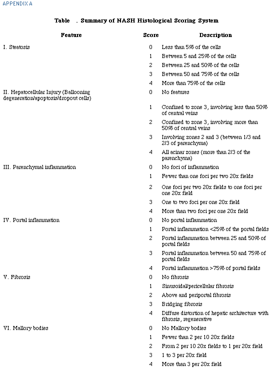
Liver biopsies were obtained using a 16-gauge Klatskin needle. A liver specimen of 15 mm with at least 10 portal tracts was considered adequate for evaluation. After conclusion of the study, all liver biopsies samples were coded and read by a hepatic pathologist (D.E.K) without the knowledge of the patient or the sequence of the biopsy. Six histological features of NASH were scored semiquantitatively from 0 to 4, including steatosis, acinar zone 3 hepatocellular injury (ballooning degeneration), parenchymal inflammation, portal inflammation, perisinusoidal fibrosis, and Mallory bodies (see Appendix A). Ubiquitin immunostaining was used to help identify Mallory bodies. Results were compared to the reading and scoring made in an unblinded fashion at the time of the biopsy. Intraobserver agreement between two readings was good to excellent with statistics ranging from 0.72 to 0.94.
The primary outcome measure for this study was improvement in liver histology as assessed by the NASH activity index. The NASH activity index was defined by the sum of scores for steatosis, parenchymal inflammation, and hepatocellular injury, and thus ranged from 0 to 12. Improvement was defined as a decrease in the NASH activity index of at least 3 points with improvements of at least 1 point for each of the three features.
Results
Patients Enrolled
Between March 2001 and April 2002, 25 patients suspected of having NASH were evaluated, and 18 were enrolled in the study. Reasons for exclusion included patient unwillingness to undergo liver biopsy (n = 2); normalization of aminotransferase levels during the lead-in phase (n = 2); development of fasting hyperglycemia, documented by fasting glucose 126 mg/dL on two separate occasions (n = 1); liver biopsy showing steatosis without inflammation and necrosis (n = 1); and a complicating intercurrent illness (n = 1).
The 18 enrolled patients were either overweight or obese: Overweight, BMI 25-29 kg/m2--7 (39%). Serum ALT levels ranged from 43 to 325 U/L and averaged 99 U/L. No enrolled patient had fasting hyperglycemia, although two had abnormal glucose tolerance with 2-hour glucose levels in the diabetic range (207 and 289 mg/dL). Liver histology showed NASH in all patients, with the NASH activity index ranging from 3 to 11. No patient had cirrhosis, but 89% had fibrosis, and 83% had Mallory's hyaline. The median duration from baseline liver biopsy to initiation of pioglitazone was 49.5 days (range, 6-297 days).
Biochemical Responses
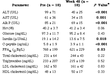
During therapy, serum ALT levels decreased in all patients, and after 48 weeks, both ALT and AST levels were in the normal range in 13 patients (72%). Serum ALT levels fell from an average of 99 U/L at baseline to 40 U/L at 48 weeks (P < 0.01). Similar improvements occurred in serum AST and alkaline phosphatase values. The ALT decreases were gradual, beginning after 4 weeks of treatment, and reaching
lowest levels between 40 and 48 weeks. Serum glucose levels did not change significantly, but indices of insulin resistance improved, including mean fasting insulin (21% decrease, P = 0.02) and C-peptide levels (29% decrease, P < 0.01). Free fatty acid levels decreased slightly (16% from baseline, P = 0.03). There were no significant changes in total cholesterol, triglycerides, LDL and HDL cholesterol levels.
Table 2. Fasting Biochemical Data at Baseline and After 48 Weeks of Pioglitazone
|
|
| |
| |
 |
|
| |
| |
NOTE. Data are presented as mean ± SD. FFAm: mean free fatty acid levels from three baseline visits and lasts three visits on pioglitazone.
Abbreviations: ALT, alanine aminotransferase; AST, aspartate aminotransferase; Alk P, alkaline phosphatase; Hct, [TK from AU]; LDL, low-density lipoprotein; HDL,
high-density lipoprotein.
Histological Responses (Table 3)
Repeat liver biopsies were available on all 18 patients. Each of the component features of the NASH activity index (steatosis, parenchymal inflammation, and hepatocellular injury) improved significantly, as did fibrosis and Mallory bodies. Eleven patients (61%) had improvement in fibrosis scores (seven patients had one level reduction, and four patients had two levels reduction of fibrosis score).
Fibrosis scores were unchanged in four (22%) and increased in three patients (17%). Nine patients (50%) had bridging fibrosis at study entry. Among these nine patients, three (33%) had stable fibrosis, and six (67%) had improvement of fibrosis (four patients had stage 2, and two patients had stage 1 fibrosis at the end of the study).
|
|
| |
| |
 |
|
| |
| |
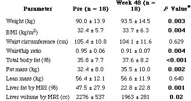
Anthropometric Results (Table 4)
A majority of patients (72%) gained weight during pioglitazone therapy. The average increase in weight was 3.5 (range, -1.7 to +13.3) kilograms, which represented a 4% increase from baseline (range, -1.7% to +12.4%). Mean body mass index increased 1.3 ± 1.7 kg/m2 (P = 0.004). Waist circumferences did not change, but waist-to-hip ratios decreased significantly (P = 0.004). Whole body composition by DEXA showed that the weight gain was mostly due to an increase in adiposity, with significant increases in total fat mass (+3.1 ± 3.6 kg, P = 0.002), but little or no change in lean body mass (+0.2 ± 2.2 kg, P = not significant). Weight gain was highly correlated with increase in total fat mass by DEXA ([r];=0.93, P < 0.001). In contrast, liver fat and liver volume as assessed by MRI both decreased significantly. There was a strong positive correlation between changes of fat in the liver as determined by MRI and degrees of steatosis by liver biopsy (r = 0.73,
P = 0.003).
Table 4. Anthropometric Data at Baseline and After 48 Weeks of Pioglitazone
|
|
| |
| |
 |
|
| |
| |
Oral Glucose Tolerance Test (Table 5)
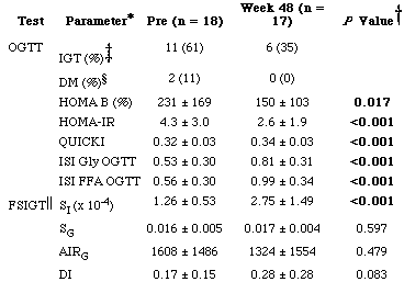
At baseline, 11 (61%) patients had impaired glucose tolerance, and two (11%) had values consistent with diabetes (2-hr glucose values 200 mg/dL). After treatment, only six subjects had impaired glucose tolerance, and no patient had 2-hr postglucose load values in the diabetic range. Insulin sensitivity indices as determined by homeostasis model assessment of insulin resistance (HOMA-IR) and quantitative insulin sensitivity check index (QUICKI) improved significantly after treatment (P < 0.001). -cell secretion as determined by homeostatic model assessment of pancreatic beta-cell function (HOMA B) percentage decreased by 77 ± 120% (P = 0.02), and insulin sensitivity index for free fatty acids derived from the OGTT also improved (P < 0.0001).
Table 5. Insulin Sensitivity Data at Baseline and After 48 Weeks of Pioglitazone
|
|
| |
| |
 |
|
| |
| |
Frequently Sampled Intravenous Glucose Tolerance Test (Table 5)
Insulin sensitivity index (SI) improved uniformly in all 17 patients from whom values were available (FSIGT was technically inadequate in one patient). The mean increase of the insulin sensitivity index was 126 ± 78% after 48 weeks of treatment. Glucose effectiveness (SG) did not change significantly, nor did the acute insulin response to glucose (AIRG), the disposition index (DI), or glucose disappearance (KG) rates (data not shown).
Side Effects and Adverse Events.
Pioglitazone was generally well tolerated. No patient developed worsened serum ALT levels. Seventeen patients completed 48 weeks of therapy without dose reduction or discontinuation. One patient discontinued therapy after 3 weeks because of dizziness. Blood glucose levels were not documented during symptomatic episodes and were normal when tested later. This patient continued to be followed in the study, underwent repeat evaluation and liver biopsy, and was included in the analysis on the basis of intention-to-treat.
Weight gain was the major side effect of therapy, averaging 3.5 kg and occurring in 72% of patients. No patient developed anemia or clinically apparent edema, reported side effects of pioglitazone. One patient had a myocardial infarction during therapy, and another suffered recurrent autoimmune uveitis during the study period. Both adverse reactions were believed to be unrelated to pioglitazone therapy, and both patients continued to take pioglitazone during these episodes.
Discussion by authors
The mechanisms of action of the TZDs are not fully understood. The TZDs improve insulin resistance by increasing glucose disposal in muscle and decreasing hepatic glucose output.[26] The agents are agonists for the peroxisome proliferator-activated receptor-gamma (PPAR), a nuclear receptor expressed in adipose tissue, muscle, and the liver. This nuclear receptor is expressed at the highest level in adipocytes, where it promotes cell differentiation[27] and decreases lipolysis and free fatty acid release.[28] The TZDs, therefore, may act by redistributing lipid from muscle and liver to peripheral adipocytes.[29] The results of this clinical study support this view, in that most patients had a decrease in liver fat, but an increase in total body adiposity. By redistributing fat from the liver to the periphery, pioglitazone may act to reduce hepatic steatosis and susceptibility to concurrent hepatocellular injury (the second hit). Another TZD, rosiglitazone has
been shown to reduce hepatic fat content and improve biochemical evidence of hepatic inflammation in patients with NASH.[18]
Using strict predetermined criteria, histological improvement was identified in two-thirds of treated patients. Notably, the degree of histological improvement was not reliably reflected by changes in serum aminotransferase levels. Thus, 83% of patients with a histological response had normal ALT levels, but so did 50% of patients who did not achieve a histological response. These results underscore the
importance of hepatic histology as an endpoint for trials of therapy in NASH. Serum ALT levels have been reported to improve with several medical therapies of NASH, including vitamin E, ursodiol, and metformin. Without histological confirmation of improvement, serum ALT improvements cannot be considered reliable in reflecting the efficacy of therapy.
A major limitation of this study was the lack of a concurrently followed control group. The natural progression of hepatic histology in NASH is not well defined. A recent placebo-controlled trial of ursodiol in NASH found that a proportion of patients receiving placebo had decreases in steatosis, but that improvements in inflammation and cell injury were uncommon.[30] Importantly, the histological improvements described in the current study involved all of the major components of NASH, including steatosis, cellular injury, parenchymal inflammation, Mallory's hyaline, and fibrosis, a combination of features that have not been found to improve spontaneously. Furthermore, the degrees of improvements in NASH activity scores were marked. At the end of treatment, many patients no longer satisfied minimal histological criteria for the diagnosis of definite NASH. Thus, the improvements seen were not simple decreases in amount of steatosis, but were histological remissions in disease. Improvements in fibrosis scores also occurred, suggesting that resolution of cell injury might eventually lead to resolution of fibrosis.
Another important shortcoming of this study was that patients were treated for 48 weeks only. Thus, it is unclear whether the biochemical and histological improvements can be sustained long-term. Furthermore, an important side effect of therapy was weight gain, which averaged 3.5 kg. Patients with NASH are usually overweight or obese, and therapies that lead to further weight gain must be viewed with caution. Continued therapy with pioglitazone may lead to continued weight gain, which might eventually reverse any beneficial effect on hepatic steatosis. For these reasons, long-term studies that support the continued efficacy and safety of the TZDs or other insulin sensitizing agents in NASH are needed before these medications can be recommended as routine therapy for NASH.
The marked increase in overweight and obesity in the Western world during the last two decades has major implications for public health. While the cardiovascular and diabetic complications of obesity have been the focus of most attention, the possible hepatic effects of increased body weight are also important and have brought NASH to increasing medical attention. Prospective studies of medical therapy of NASH are important in elucidating its pathogenesis and developing means of ameliorating or preventing injury.
|
|
| |
| |
 |
|
| |
| |
|
|
| |
| |
 |
|
| |
| |
1 Bacon BR, Farahvash MJ, Janney CG, Neuschwander-Tetri BA. Nonalcoholic steatohepatitis: an expanded clinical entity. Gastroenterology 1994; 107: 1103-1109. Links
2 Ludwig J, Viggiano TR, McGill DB, Oh BJ. Nonalcoholic steatohepatitis: Mayo Clinic experiences with a hitherto unnamed disease. Mayo Clin Proc 1980; 55: 434-438. Links
3 Matteoni CA, Younossi ZM, Gramlich T, Boparai N, Liu YC, McCullough AJ. Nonalcoholic fatty liver disease: a spectrum of clinical and pathological severity. Gastroenterology 1999; 116: 1413-1419. Links
4 Ruhl CE, Everhart JE. Determinants of the association of overweight with elevated serum alanine aminotransferase activity in the United States. Gastroenterology 2003; 124: 71-79. Links
5 Powell EE, Cooksley WG, Hanson R, Searle J, Halliday JW, Powell LW. The natural history of nonalcoholic steatohepatitis: a follow-up study of forty-two patients for up to 21 years. HEPATOLOGY 1990; 11: 74-80. Links
6 Wanless IR, Lentz JS. Fatty liver hepatitis (steatohepatitis) and obesity: an autopsy study with analysis of risk factors. HEPATOLOGY 1990; 12: 1106-10. Links
7 Teli MR, James OF, Burt AD, Bennett MK, Day CP. The natural history of nonalcoholic fatty liver: a follow-up study. HEPATOLOGY 1995; 22: 1714-1719. Links
8 Caldwell SH, Oelsner DH, Iezzoni JC, Hespenheide EE, Battle EH, Driscoll CJ. Cryptogenic cirrhosis: clinical characterization and risk factors for underlying disease. HEPATOLOGY 1999; 29: 664-669. Links
9 Bugianesi E, Leone N, Vanni E, Marchesini G, Brunello F, Carucci P, Musso A, et al. Expanding the natural history of nonalcoholic steatohepatitis: from cryptogenic cirrhosis to hepatocellular carcinoma. Gastroenterology 2002; 123: 134-140. Links
10 James O, Day C. Non-alcoholic steatohepatitis: another disease of affluence. Lancet 1999; 353: 1634-1636. Links
11 Day CP. Non-alcoholic steatohepatitis (NASH): where are we now and where are we going? Gut 2002; 50: 585-588. Links
12 Sanyal AJ, Campbell-Sargent C, Mirshahi F, Rizzo WB, Contos MJ, Sterling RK, Luketic VA, et al. Nonalcoholic steatohepatitis: association of insulin resistance and mitochondrial abnormalities. Gastroenterology 2001; 120: 1183-1192. Links
13 Marchesini G, Brizi M, Bianchi G, Tomassetti S, Bugianesi E, Lenzi M, McCullough AJ, et al. Nonalcoholic fatty liver disease: a feature of the metabolic syndrome. Diabetes 2001; 50: 1844-1850. Links
14 Diabetes Prevention Program Research Group. Reduction in the incidence of type 2 diabetes with lifestyle intervention or metformin. N Engl J Med 2002; 346: 393-403. Links
15 Lin HZ, Yang SQ, Chuckaree C, Kuhajda F, Ronnet G, Diehl AM. Metformin reverses fatty liver disease in obese, leptin-deficient mice. Nat Med 2000; 6: 998-1003. Links
16 Marchesini G, Brizi M, Bianchi G, Tomassetti S, Zoli M, Melchionda N. Metformin in non-alcoholic steatohepatitis. Lancet 2001; 358: 893-894. Links
17 Caldwell SH, Hespenheide EE, Redick JA, Iezzoni JC, Battle EH, Sheppard BL. A pilot study of a thiazolidinedione, troglitazone, in nonalcoholic steatohepatitis. Am J Gastroenterol 2001; 96: 519-525. Links
18 Neuschwander-Tetri BA, Brunt EM, Wehmeier KR, Oliver D, Bacon BR. Improved nonalcoholic steatohepatitis after 48 weeks of treatment with the PPAR- ligand rosiglitazone. HEPATOLOGY 2003; 38: 1008-1017. Links
19 Sanyal AJ, Contos MJ, Sargeant M, Stravitz RT, Luketic VA, Sterling RK, Shiffman ML, et al. A randomized controlled pilot study of pioglitazone and vitamin E versus Vitamin E for nonalcoholic steatohepatitis [abstract]. HEPATOLOGY 2002; 36( suppl): 875A. Links
20 The Food Guide Pyramid. Washington, DC: Department of Agriculture, Center for Nutrition Policy and Promotion; 1996. Home and Garden Bulletin No. 252.
21 Lohman TG, Roche AF, Martorell R. Anthropometric Standardization Manual. Champaign, IL: Human Kinetics Publishers, Inc., 1988: 1-177.
22 World Health Organization. Diabetes Mellitus: Report of a WHO Study Group. Technical Report Series, No. 727. Geneva: World Health Organization, 1985; 1-113.
23 Bergman RN. Lilly lecture 1989. Toward physiological understanding of glucose tolerance: minimal-model approach. Diabetes 1989; 38: 1512-1527. Links
24 Fishbein MH, Gardner KG, Potter CJ, Schmalbrock P, Smith MA. Introduction of fast MR imaging in the assessment of hepatic steatosis. Magn Reson Imaging 1997; 15: 287-293. Links
25 Levenson H, Greensite F, Hoefs J, Friloux L, Applegate G, Silva E, Kanel G, et al. Fatty infiltration of the liver: quantification with phase-contrast MR imaging at 1.5 T vs biopsy. AJR Am J Roentgenol 1991; 156: 307-312. Links
26 Inzucchi SE, Maggs DG, Spollett GR, Page SL, Rife FS, Walton V, Shulman GI. Efficacy and metabolic effects of metformin and troglitazone in type II diabetes mellitus. N Engl J Med 1998; 338: 867-873. Links
27 Okuno A, Tamemoto H, Tobe K, Ueki K, Mori Y, Iwamoto K, Umesono K, et al. Troglitazone increases the number of small adipocytes without the change of white adipose tissue mass in obese Zucker rats. J Clin Invest 1998; 101: 1354-1361. Links
28 Guan HP, Li Y, Jensen MV, Newgard CB, Steppan CM, Lazar MA. A futile metabolic cycle activated in adipocytes by antidiabetic agents. Nat Med 2002; 8: 1122-1128. Links
29 Mayerson AB, Hundal RS, Dufour S, Lebon V, Befroy D, Cline GW, Enocksson S, et al. The effects of rosiglitazone on insulin sensitivity, lipolysis, and hepatic and skeletal muscle triglyceride content in patients with type 2 diabetes. Diabetes 2002; 51: 797-802. Links
30 Lindor KD. UDCAsol;NASH Study Group. Ursodeoxycholic acid for treatment of nonalcoholic steatohepatitis: results of a randomized, placebo-controlled trial [abstract]. Gastroenterology 2003; 124( suppl): 336A. Links
31 Executive Summary of The Third Report of The National Cholesterol Education Program (NCEP) Expert Panel on Detection, Evaluation, and Treatment of High Blood Cholesterol in Adults (Adult Treatment Panel III). JAMA 2001; 285: 2486-2497. Links
32 Matthews DR, Hosker JP, Rudenski AS, Naylor BA, Treacher DF, Turner RC. Homeostasis model assessment: Insulin resistance and beta-cell function from fasting plasma glucose and insulin concentrations in man. Diabetologia 1985; 28: 412-419. Links
33 Katz A, Nambi SS, Mather K, Baron AD, Follmann DA, Sullivan G, Quon MJ. Quantitative insulin sensitivity check index: a simple, accurate method for assessing insulin sensitivity in humans. J Clin Endocrinol Metab 2000; 85: 2402-2410. Links
34 Nair S, Diehl AM, Perrillo R. Metformin in nonalcoholic steatohepatitis (NASH): efficacy and safety, a preliminary report [abstract]. Gastroenterology 2002; 122( Suppl): 4A. Links
35 Fuller NJ, Laskey MA, Elia M. Assessment of the composition of major body regions by dual-energy X-ray absorptiometry (DEXA), with special reference to limb muscle mass. Clin Physiol 1992; 12: 253-266. Links
|
|
| |
| |
|
|
|