| |
Review article: drug therapy for non-alcoholic fatty liver disease
|
| |
| |
".....the focus of treatments should be on correction of underlying metabolic syndrome...."
Alimentary Pharmacology & Therapeutics
Volume 23 Page 207 - January 2006
K. M. COMAR & R. K. STERLING
Department of Internal Medicine, Division of Gastroenterology, Hepatology and Nutrition, Virginia Commonwealth University, Richmond, VA, USA
Table 3. List of drugs that have been or are being studied for the treatment of NAFLD
Insulin sensitizers
Metformin
Pioglitazone
Rosiglitazone
Antioxidants
Vitamin E
Vitamin C
Hepatoprotective agents
Betaine
Ursodeoxycholic acid
Pentoxyfylline
Summary
Non-alcoholic fatty liver disease represents a spectrum of liver diseases, characterized mainly by macrovesicular steatosis in the absence of significant alcohol ingestion. Non-alcoholic fatty liver disease includes both non-alcoholic fatty liver and non-alcoholic steatohepatitis.
Non-alcoholic steatohepatitis once considered a benign process is now known to lead to progressive fibrosis and cirrhosis. Histologically indistinguishable from alcoholic liver disease, the exact aetiology of non-alcoholic fatty liver disease remains unknown, but the fundamental pathophysiological process appears to be insulin resistance and oxidative stress related to the metabolic syndrome.
Therapy has focused on risk factors, weight reduction and pharmacological intervention. Promising pharmacological treatments have been demonstrated with antioxidants, insulin sensitizers, hepatoprotectants and lipid-lowering agents. However, without larger randomized studies, no pharmacological treatments can be recommended at this time.
Introduction
Non-alcoholic fatty liver disease (NAFLD) represents a spectrum of liver diseases characterized mainly by macrovesicular steatosis in the absence of significant alcohol ingestion. The hepatic histology can vary from steatohepatitis to isolated hepatic steatosis, referred to as non-alcoholic steatohepatitis (NASH) or its precursor non-alcoholic fatty liver disease (NAFL), respectively. NASH once considered a benign process has been found to lead to progressive fibrosis and cirrhosis.14 The transition of NAFL to NASH is not clearly demarcated; however, histologically, NASH will show the presence of cytological ballooning, Mallory's hyaline, scattered inflammation and pericellular fibrosis.5 Because the presence of inflammation and fibrosis is distinctive features in NASH, a liver biopsy is often needed to differentiate NAFL from NASH.6
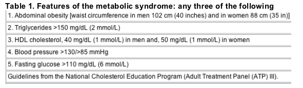
It is estimated that 12-15% of the general population has NAFL while 3-4% have NASH.7 While a majority of cases occur in females between the ages of 40 and 60, reports in children are well documented. NAFLD association with the metabolic syndrome is also well documented (Table 1).8 All subjects with the metabolic syndrome are potentially at risk for having NAFLD. An estimated 70-80% of obese individuals have NAFL while 15-20% have NASH.9 Approximately 50-100% of subjects with NASH are overweight,102 50-60% are hypertensive and 50-60% have dyslipidemias. While the exact aetiology of NAFLD remains unknown, the fundamental pathophysiologic process that connects the diverse conditions associated with NAFLD seems to be insulin resistance.12 Insulin resistance is present in approximately 98% of individuals with NAFLD and over 80% of subjects meet the criteria for the metabolic syndrome.13
Evaluation
The ideal conditions for screening for a disease include high prevalence, availability of non-invasive diagnostic screening tools and the ability to provide specific effective interventions. Over 47 million individuals in the US are estimated to have the metabolic syndrome and an estimated 80% of such individuals have features of NAFLD.14, 15 It is therefore safe to say that the prevalence of NAFLD is clearly quite high. However, the diagnostic accuracy of non-invasive tools remains poor and, moreover, there is no clear-cut established pharmacologic treatment for NAFLD. Thus, the data do not support the routine screening of asymptomatic individuals with metabolic syndrome for NAFLD.
Symptoms of NAFLD include non-specific findings such as abnormal liver enzymes, right upper quadrant discomfort, unexplained hepatomegaly or the incidental findings of fatty liver disease on abdominal imaging.16 However, in an analysis of 325 patients with NASH and 75 with NAFLD alone at our centre, these features were similar between groups and were not able to differentiate these conditions (unpublished data, VCUHS). Liver aminotransamenase is elevated in close to 90% of patients with NASH.10 Typically, aspartate aminotransferase (AST) as well as alanine aminotransferase (ALT) values are under 250 IU/L and unlike alcoholic liver disease an AST/ALT ratio is of less than 1. An ALT or AST value >300 IU/L should raise suspicion of alternate pathology. Alkaline phosphatase (ALP) will be elevated in up to half of the affected individuals, usually not exceeding two to three times the upper limit of normal values. Unfortunately, the clinical and laboratory findings while suggestive of NAFLD are also typical in other causes of liver disease. Moreover, the histopathology in NASH is indistinguishable for the alcoholic liver disease. It is therefore critically important to make an accurate diagnosis in order to optimize management strategies.
Diagnosis
The principle objectives in patients with suspected NAFLD are confirmation of the diagnosis, identification of aetiology, evaluation of prognosis and management of disease. The gold reference standard for the diagnosis of fatty liver disease is a liver biopsy. However, as with chronic hepatitis C, it is subject to sampling error and both intra- and interobserver variability.17 In addition, the risks and costs associated with liver biopsies with the lack of therapeutic options have led to a considerable interest in non-invasive diagnostic methods. Currently, hepatic sonogram is the most commonly used imaging modality in the diagnosis of fatty liver. Sonographic features of NAFLD include the increased hepatic parenchymal echo texture and vascular blurring.9, 18, 19 Unfortunately, these findings are also seen in other forms of liver disease, and the ability to detect fatty liver by sonography diminishes markedly once the degree of hepatic steatosis decreases to 30% or less.20 Joseph et al. found ultrasound to have 89% sensitivity and 93% specificity with a positive likelihood ration of 1221 and a negative likelihood ration of 0.12 for steatosis. Therefore, using Fagan's nomogram,22 the positive and negative predictive values for the presence or absence of steatosis on ultrasound in a patient with abnormal liver tests in the absence of other causes of hepatitis are 96% and 19%, respectively.23 Computerized tomography (CT) and magnetic resonance imaging (MRI) provide another diagnostic option, but the modest increase in diagnostic accuracy is at the expense of a marked increase in cost.
Aetiology
In the presence of features of the metabolic syndrome, further evaluation of NAFLD aetiology is often unwarranted. It should however be noted that NAFLD can also be caused by specific metabolic or iatrogenic conditions distinct from the metabolic syndrome. (Table 2) The evaluation in a patient with non-metabolic syndrome associated NAFLD should consist of a detailed alcohol, drug and surgical history. Assessment of adipose tissue distribution and fasting lipid profile can help in identifying a lipodystrophy or rare metabolic syndromes. It is generally believed that a fatty liver does not develop with alcohol consumption levels <20 g/day.
Table 2. Causes of NAFLD
The metabolic syndrome
Drugs
Corticosteroids
Tamoxifen
Amiodarone
Diltiazem
Protease inhibitors
Nucleoside reverse transcriptase inhibitors
Lipodystrophy (fat redistribution syndromes)
Congenital vs. acquired (HIV-related)
Lipid loss vs. lipid deposition syndromes
Rare metabolic syndromes
Abetalipoproteinaemia
Hypobetalipoproteinaemia
Weber-Christian syndrome
Total parenteral nutrition
Short bowel syndrome
Small bowel bacterial overgrowth, e.g. small bowel diverticulosis
Chronic inflammatory disorders, e.g. rheumatoid arthritis and systemic lupus erythematosus
Prognosis
Prognosis of a patient with NAFLD depends on histology, clinical features and the presence of additional morbidities. The presence of NASH vs. NAFL and the stage of fibrosis provide a gradient for prognosis. Signs of liver decompensation, such as ascites, varices, hepatic encephalopathy, hypoalbuminaemia, hyperbilirubinaemia as well as prothrombin time, provide clinical and laboratory markers for prognosis. As with any patient, prognosis depends and should take into account additional medical problems. Patients with underlying metabolic syndrome are especially at risk for cardiovascular and cerebral vascular morbidity and mortality. A recent study of 420 patients with NAFLD in the community setting found both liver related and overall mortality higher than the general population and associated with older age, impaired fasting glucose and cirrhosis.24
Management
Therapeutic interventions are limited in NASH. Therapy has focused on modifications of risk factors, weight reduction and pathogenesis-oriented pharmacological therapies. Weight loss remains the mainstay in non-pharmacological intervention available in NASH patients. Weight loss in overweight patients with liver disease has shown the sustained improvement in liver enzymes.25 In addition, improvement or resolution of obesity and metabolic syndrome has associated major improvements in lobular steatosis, necroinflammatory changes and fibrosis.26
Gradual weight reduction and increased physical activity in overweight patients with liver disease have not only shown improvement in liver enzymes, but also serum insulin levels, and quality of life.25 In addition, weight loss after surgery has also demonstrated major improvement in abnormal liver histological features found in severely obese patients.26 While no particular type of diet has been shown to be superior to another, pending comorbidities a standard diabetic or healthy heart diet should be recommended. Patient should be advised to aim to lose 5-10% of their baseline weight at a rate of 500 g-1.5 kg/week. Rapid weight loss should be advised against secondary to risks of exacerbating liver damage.
The effects on pharmacological therapy unlike weight loss have unfortunately not been as clear-cut. The absence of empowered, randomized, controlled trials with histological endpoints and long-term morbidity/mortality outcomes make it difficult to advocate specific treatments. The existing literature and on-going studies have focused on four pharmacological therapies: (i) antioxidants, (ii) insulin sensitizer, (iii) hepatoprotectants and (iv) lipid-lowering agents. This review will examine the studies that have evaluated the potential pharmacological modalities in patients with NASH, their effectiveness and limitations (Table 3).
Table 3. List of drugs that have been or are being studied for the treatment of NAFLD
Insulin sensitizers
Metformin
Pioglitazone
Rosiglitazone
Antioxidants
Vitamin E
Vitamin C
Hepatoprotective agents
Betaine
Ursodeoxycholic acid
Pentoxyfylline
Vitamins E and C
While the exact pathogenesis of NASH still remains an enigma, oxidative stress is believed to be a key catalyst in the development of NASH. Oxidation of cytotoxic-free acids in rat models has shown to induce liver cyotchrome P450 enzymes and deplete hepatic antioxidant concentrations.27 In addition, by-products of oxidative stress like 4-hydroxynonenal (HNE) have been shown to be involved in excess extracellular matrix deposition.28
It would therefore be natural to presume that antioxidants such as vitamins E and C would help protect against the damaging effects of free radicals in the liver. Multiple studies have evaluated the potential of vitamins in the treatment if NASH. An open-label pilot study by Lavine et al. evaluated vitamin E in the obese children. These children had chronically elevated serum aminotransferase secondary to NASH. They were given vitamin E (400-1200 IU daily) and monitored for 4-10 months. The body mass index of the children did not change significantly with treatment; however, a significant decline in aminotransferase was noted.29 Of the 11 subjects enrolled in the study, ALT decreased from 175 to 40 IU/L (P < 0.002), AST decreased from 104 to 33 IU/L (P < 0.002), and ALP decreased from 279 to 202 IU/L (P < 0.003).29 Enzyme levels not only remained normal during the treatment, but also returned to abnormal levels in those electing to terminate treatment.
In an additional small study by Hasegawa et al., 12 patients were given dietary instructions for 6 months, and then alpha-tocopherol (300 mg/day) for 1 year. Blood chemistries, measurement of plasma transforming growth factor-beta 1 and liver biopsies were undertaken before and after the 1-year alpha-tocopherol treatment. The study found that histological findings, such as steatosis, inflammation and fibrosis, in NASH patients improved after alpha-tocopherol treatment.30
Prospective randomized studies have also shown improvement of fibrosis in NASH with vitamin E.31, 32 Harrison et al. in a prospective, double-blind, randomized, placebo-controlled trial with 45 patients showed that 6 months of vitamins E and C combination (1000 IU and 1000 mg, respectively) resulted in significant improvement in fibrosis score (P = 0.002).31 In addition, Sanyal et al. has demonstrated that vitamin E, when combined with an insulin sensitizer (pioglitazone), produced a significant decrease in steatosis (mean 2 vs. 1; P < 0.002).32 Despite these promising results in pilot studies, vitamin E is not recommended outside of clinical trials in patients with NASH because a meta-analysis of high-dose vitamin E supplementation has shown to increase all-cause mortality.33
Betaine
Histological findings of NASH and alcoholic disease are often indistinguishable. Betaine, a component of the metabolic cycle of methione, has been shown to increase S-adenosylmethionine levels (SAM) and protect against steatosis in animal models of alcoholic liver disease.34 The efficacy of betaine in patients with NASH was demonstrated in a pilot study involving 10 adult patients. Barack et al. demonstrated that seven out of 10 patients who completed 1 year of betaine treatment were found to have a significant improvement in serum levels of ALT (P = 0.02), AST (P = 0.007), degree of steatosis, necroinflammatory grade and stage of fibrosis.34
Metformin
There is a clear-cut relationship between fatty liver disease and hyperinsulinemic insulin resistance in patients.3436 On the basis of this relationship, it has been postulated that a decrease in insulin resistance may be of therapeutic value in fatty liver disease. Metformin, a biquanide, reduces hyperinsulinaemia and improves hepatic insulin resistance.34, 3739 Its major site of action appears to be in the mitochondria, and it has been shown to stimulate pyruvate-kinase,40 fatty acid beta-oxidation, anaerobic respiration (i.e. lactate production)41 as well as suppress the expression of lipogenic enzymes.42
In insulin-resistant ob/ob mice with fatty liver disease, metformin improved fatty liver disease, reversed hepatomegaly, steatosis and aminotransferase abnormalities.43 Nevertheless, in human trials, metformin has shown mixed results. In one uncontrolled pilot study, compliant individuals on long-term metformin had significantly reduced mean transaminase concentrations when compared with non-complaint patients.44 In another open-label pilot study, 15 patients with NASH who completed 1 year of treatment of metformin (20 mg/kg) showed during the initial 3 months an improvement in ALT, AST (P-value = 0.01 and 0.02, respectively) and insulin sensitivity during the initial 3 months. However, after 3 months, there was no further improvement in insulin sensitivity and there was a gradual rise in AST and ALT back to pre-treatment levels.45
In a controlled trial, Uygun et al. randomized 36 patients with NASH. These patients were divided into two groups: a dietary treatment group and a dietary treatment plus metformin for 6-month group.46 In the metformin/diet group vs. diet alone group, a significant decrease in the mean ALT (U/L) 37 vs. 17 (P = 0.003), AST (U/L) 22 vs. 7 (P = 0.0001), insulin (IU/mL) 5 vs. 2 (P = 0.002) and C-peptide (ng/mL) 1 vs. 0.1 (P = 0.002) was noted. In addition to biochemical markers, an improvement in necroinflammation was also observed in the metformin group but the results did not achieve statistical significance.46
While initial studies of metformin have been promising, long-term benefits have yet to be clarified. There are currently two large clinical trials (PIVENS and TONIC) that are being initiated by the National Institutes of Health to address this issue. Until the results of these trials are published, these agents should not be used in routine clinical practice for non-diabetic patients with NASH.
Angiotensin-converting enzyme inhibitors
The renin-angiotensin system (RAS) is frequently activated in patients with chronic liver disease. In animal models, evidence has shown that angiotensin 2 receptor antagonist and angiotensin-converting enzyme (ACE) inhibitors display antifibrotic characteristics via the hepatic stellate cell proliferation.47 In a pilot study examining the therapeutic efficacy of angiotensin 2 receptor antagonist, losartan was studied in patients with NASH and hypertension. Seven patients were treated with losartan (50 mg/day) for 48 weeks. After 48 weeks, patients not only showed a significant decrease in blood markers of hepatic fibrosis, but also an improvement in serum aminotransferase levels. However, because the small number of patients in this study limits the validity, a larger-controlled study is needed.
Urodeoxycholic acid
Ursodeoxycholic acid (UDCA) is a bile acid, which has various cytoprotective, antiapoptotic and immunomodulatory properties.48, 49 The UDCA decreases levels of endogenous hydrophobic bile acids while increasing the fraction of non-toxic hydrophilic bile acids. Hepatoprotective properties have lead to UDCA being used to treat liver diseases such as primary biliary cirrhosis (PBC), primary sclerosing cholangitis (PSC) and cystic fibrosis-related cholestasis. An initial pilot study had shown significant decrease in mean ALP, ALT, GGT and grade of hepatic steatosis after 1 year of UDCA at dose of 13-15 mg/kg/day.50 On the contrary, a larger placebo-controlled randomized trial showed that 2 years of UDCA therapy at similar doses, while safe and well-tolerated did not show changes in the degree of steatosis, necroinflammation or fibrosis.51 Therefore, its routine use in NASH cannot be recommended.
Pentoxifylline
The production of tumour necrosis factor-alpha (TNF-alpha) is one of the primary events in many types of liver injury. TNF-alpha triggers the production of additional cytokines that collectively recruit inflammatory cells, which destroy hepatocytes and induce fibrogenesis.52 Patients with NASH have been shown to have higher levels of TNF-alpha.53, 54 Pentoxifylline (PTX) is a methylxanthine compound known to inhibit the production of TNF-alpha.55 Two pilot studies have evaluated the possible role of PTX in the treatment of NASH.
Both studies consisted of histologically proven NASH, in 20 patients or less, and treatment ranged from 6 months to 1 year. Biochemical improvement was demonstrated in each study; however, histological follow-up was not obtained and gastrointestinal side effects lead to a high rate of withdrawal.56
Thiazolidninediones
Thiazolidinediones (TZDs) are a class of antidiabetic agents that increase insulin sensitivity in peripheral adipocytes. TZDs lower plasma fatty acid concentrations and redistribute intracellular lipids.57 Oral TZD also reduces extracellular matrix deposition and hepatic stellate cells activation in both toxic and cholestatic models of liver fibrosis.58 Three members in the TZD class have been evaluated in NASH patients: troglitazone, rosiglitazone and proglitazone.
Troglitazone
Troglitazone was used in a study with 10 female patients (mean age 44) with histological NASH. Troglitazone was given at a dose of 400 mg/day for < or = 6 months. Seven of the 10 patients responded with normal ALT at the end of the treatment. In the responders, ALT fell from 87 to 39 by the end of treatment (P = 0.01), and AST decreased from 77 to 30 (P = 0.002). Histological comparisons, before and after therapy, showed persistent steatohepatitis with only mild improvement.59 However, recent compelling data have shown troglitazone to be associated with acute idiopathic hepatitis and currently have been withdrawn from the market.60, 61
Rosiglitazone
Thirty adults with prior biopsy evident for NASH were enrolled to receive rosiglitazone, 4 mg b.d. for 48 weeks. All patients were overweight and had impaired glucose tolerance or diabetes. Of the 25 patients who completed the trial, significant improvement in insulin sensitivity and mean serum ALT levels (104 initially, 42 U/L at the end of treatment) were noted.62 In addition, significant improvement in hepatocellular ballooning and perisinusoidal fibrosis also occurred. Side effects reported included a drop in haemoglobin, weight gain and bad dreams. In addition, it is often advised to use caution when using TZD in diabetics with mild elevations in liver enzymes and is often believed to be contraindicated in those with ALTs > 3 upper limit normal. Despite these beliefs, studies have suggested that diabetics with elevated baseline liver enzymes do not have a higher risk of hepatotoxicity with rosiglitazone. In fact, Chalisani et al. performed as study consisting of two cohorts of patients, one with elevated baseline liver enzymes (AST > 40 IU/L and/or ALT > 35 IU/L) and one without. Liver biochemistries were followed and evaluated over a 12-month period on rosiglitazone. The multicentre study found no higher incidence of mild-to-moderate (10% vs. 7%, P = 0.2) or -severe elevations (0.9% vs. 0.6%, P = 0.9) in liver biochemistries between the two cohorts. In fact, the frequency of discontinuing rosiglitazone therapy during the follow-up was similar between the two cohorts (9% vs. 8%, P = 1.0).63
Piogitlazone
Eighteen non-diabetic patients with biopsy-proven NASH were treated with pioglitazone (30 mg daily) for 48 weeks. For the duration of therapy, serum ALT levels decreased in all patients, and at completion both ALT and AST levels were in the normal range in 13 patients (72%).64 ALT levels decreased an average of 50 U/L from baseline. Histological features of steatosis, cellular injury, parenchymal inflammation, Mallory bodies and fibrosis were also significantly improved from baseline (all P < 0.05). Generally, well-tolerated, weight gain was the major side effect of therapy. To add, there were no reports of patients having a drop in haemoglobin or worsening serum ALT levels, as reported with other TZDS.
Probucol
Probucol is a lipid-lowering drug with potent antioxidant properties that tends to accumulate in fatty tissues.65 The effectiveness of probucol in an initial open-labelled study leads to a randomized, double-blind, placebo-controlled trial, in which 30 biopsy-proven NASH patient were given probucol 500 mg/day or placebo for 6 months.66 Of the twenty-seven patients who completed the trial, there was a significant improvement in ALT levels with normalization of aminotranferases in 50% of the probucol group, but no improvement in those who received placebo treatment. The mean AST and ALT levels changed from 82 to 36 and 102 to 45 in the treatment group and from 58 to 50 and 97 to 96 in the control group, respectively. The decrease in ALT level in the treatment group when compared with the control group was significant (95% CI: 20-94 IU).
Conclusions
The ideal pharmacological agent is one that is highly effective, safe, easy to administer, easy to tolerate and inexpensive. Such an agent does not yet exist in the treatment of NASH. In the absence of adequately powered, randomized, controlled trials with histological endpoints and long-term morbidity/mortality outcomes, it is currently difficult to make evidence-based recommendations on pharmacological treatment in NASH. However, the potential benefits of multiple agents have been postulated and tested in pilot studies. Promising therapeutic results have been demonstrated with multiple agents and currently on-going larger randomized trials are underway, which will hopefully clarify the role of specific pharmacologic treatments. Until then, the focus of treatments should be on correction of underlying metabolic syndrome.
|
|
| |
| |
|
|
|