| |
HCV is Detected in Liver of HCV Antibody-Positive But HCV RNA Negative Patients, so don't assume HCV has been cleared if viral load is undetectable
|
| |
| |
"Detection of Hepatitis C Virus (HCV) RNA in the Liver of Healthy, Anti-HCV Antibody-Positive, Serum HCV RNA-Negative Patients with Normal Alanine Aminotransferase Levels"
".....the majority of healthy patients who test positive for anti-HCV antibodies and have normal ALT levels but who do not have HCV RNA detected in serum have an ongoing HCV infection because HCV RNA is detected in the liver. The epidemiological and clinical relevance of this finding should be studied in the future....."
The Journal of Infectious Diseases July 1, 2006;194:53-60
Vicente Carreno, Margarita Pardo, Juan Manuel Lopez-Alcorocho, Elena Rodriguez-Inigo, Javier Bartolome, and Inmaculada Castillo
Fundacion para el Estudio de las Hepatitis Virales, Madrid, Spain
ABSTRACT
Background. It is unknown whether hepatitis C virus (HCV) is present in the liver of anti-HCV antibody-positive patients with persistently normal alanine aminotransferase (ALT) levels and undetectable serum HCV RNA levels.
Methods. We determined the presence of genomic and antigenomic HCV RNA strands in liver biopsy specimens and peripheral blood mononuclear cell (PBMC) samples obtained from 12 anti-HCV antibody-positive patients who had normal ALT levels and who had been serum HCV RNA negative for at least 12 months, according to the results of quantitative, strand-specific, real-time reverse-transcription-polymerase chain reaction and, also, in situ hybridization of liver cells. Intrahepatic HCV RNA was cloned and sequenced.
Results.
All patients remained anti-HCV antibody positive and serum HCV RNA negative, and all had normal ALT values during follow-up (mean duration ± SD, 29.2 ± 19.8 months).
Genomic HCV RNA was detected in liver biopsy specimens obtained from 10 (83%) of 12 patients, and the antigenomic strand was detected in 10 (100%) of 10 liver biopsy specimens in which genomic HCV RNA was detected.
Results were confirmed by in situ hybridization. Intrahepatic HCV was of genotype 1b, and HCV sequencing demonstrated no cross-contamination among samples. Genomic HCV RNA was found in 6 (50%) of 12 PBMC samples, and antigenomic HCV RNA was also detected in 5 (83%) of these 6 PBMC samples.
Conclusion. HCV may persist and replicate in the liver and PBMCs of healthy, anti-HCV antibody-positive, serum HCV RNA-negative patients who have persistently normal ALT levels. These patients should be followed up, because they have an ongoing viral infection.
Hepatitis C virus (HCV) infection is a major cause of liver disease that is characterized by the presence of antibodies against HCV proteins (i.e., anti-HCV antibodies) and HCV RNA in serum [1]. During acute HCV infection, 15%-20% of individuals clear the virus, with loss of serum HCV RNA and normalization of liver-function test results occurring but with anti-HCV antibodies remaining detectable [2]. Clearance of HCV infection also occurs in individuals with chronic hepatitis C, after receipt of successful antiviral treatment [3]. All of these individuals are considered to have completely recovered from HCV infection and to have been immunized against at least the corresponding HCV strain [4]. On the other hand, there are healthy individuals with normal alanine aminotransferase (ALT) levels who test positive for anti-HCV antibodies in the absence of serum HCV RNA and who have no history of acute or chronic liver disease [5]. This situation could reflect an immunological response to an unapparent exposure to HCV without clinical consequences, rather than a clearance of a HCV infection.
Recently, a new form of HCV infection, defined as "occult HCV infection," has been described [6]. Occult HCV infection is characterized by the absence of anti-HCV antibodies and serum HCV RNA and by the presence of HCV RNA in the peripheral blood mononuclear cells (PBMCs) of most patients (70%) and in the liver of all patients. In addition, detection of HCV RNA in the PBMCs of patients with anti-HCV antibodies and normal ALT levels years after the resolution of HCV infection (either spontaneously or induced by antiviral treatment) has also been reported [7-9]. Also, the presence of intrahepatic HCV RNA after normalization of liver enzyme levels and clearance of serum HCV RNA in patients with chronic hepatitis C who responded to an antiviral treatment has been demonstrated [9, 10]. However, up to now, the possible presence of HCV RNA in the liver tissue of healthy, anti-HCV antibody-positive patients with persistently normal ALT levels and no history of acute or chronic liver disease has not been studied. In the present study, we analyzed whether anti-HCV antibody-positive, serum HCV RNA-negative patients with persistently normal ALT levels have HCV RNA (genomic HCV RNA) in the liver and, also, whether HCV is replicating by detecting the antigenomic HCV RNA.
DISCUSSION
Anti-HCV antibody-positive, serum HCV RNA-negative patients who have normal ALT levels may be considered to be patients who have cleared HCV infection, either spontaneously or after successful antiviral treatment [2, 3]. However, recent studies [7-10] have demonstrated that HCV RNA may be present in the liver or PBMCs of such patients, thus indicating an ongoing HCV infection. To our knowledge, no studies have been performed to search for the presence of HCV RNA in the liver of healthy, anti-HCV antibody-positive, serum HCV RNA-negative patients with normal ALT levels who do not have a previous history of acute or chronic liver disease.
In the present study, HCV RNA was detected in the liver biopsy specimens of 10 (83%) of 12 healthy, anti-HCV antibody-positive patients with persistently normal ALT levels and without HCV RNA detected in serum. The specificity of the results was demonstrated by sequencing data that showed the absence of cross-contamination among the samples. Furthermore, all results were confirmed by in situ hybridization. It should be noted that 8 of the 10 patients with intrahepatic HCV RNA did not have risk behaviors for HCV infection, whereas the other 2 patients had received a blood transfusion >25 years previously.
As mentioned above, previous studies [9, 10] have also reported the presence of HCV RNA in the liver of patients who cleared HCV infection after receiving successful antiviral treatment, albeit in lower percentages (2% and 27%) than were noted in our study. Other than the differences in the populations studied, several factors could explain this discrepancy. One factor could be possible HCV RNA degradation resulting from improper preservation of liver specimens, with this leading to false-negative results [15]. Although McHutchison et al. [10] analyzed positive internal controls to ensure the integrity of the RNA in order to validate their PCR results, it has been proved that amplification of positive internal controls does not assure the integrity of HCV RNA [15]. Another cause could be the different methodology employed for HCV RNA isolation as well as for HCV RNA amplification.
In addition, the antigenomic HCV RNA strand was detected in 10 of 10 liver samples that had genomic viral RNA. This result demonstrates that HCV was replicating in liver cells in the majority of patients (83%) who had no history of liver disease but who tested positive for anti-HCV antibodies in the absence of HCV RNA in serum and who presented with constantly normal ALT levels. Therefore, it should be considered that, once HCV infects the liver, complete clearance of HCV is not a frequent event, at least in untreated patients. In light of these results, anti-HCV antibody-positive, serum HCV RNA-negative patients with normal ALT levels who are considered to have cleared HCV may, in fact, have an ongoing HCV infection. Thus, healthy patients with normal ALT levels who are found to be anti-HCV antibody positive but serum HCV RNA negative during routine screening for anti-HCV antibodies (i.e., for blood donations, pregnancy, epidemiological studies, or medical checkups) should be carefully followed up.
HCV RNA was also detected in 50% of the PBMC samples obtained from our anti-HCV antibody-positive, serum HCV RNA-negative patients who had a normal ALT level. The frequency of HCV infection noted in PBMC samples in the present study agrees with that reported by Radkowski et al. [8], who found HCV RNA in uncultured PBMC samples obtained from 7 of 11 patients who also had normal ALT values and anti-HCV antibodies but who did not have detectable viral RNA in serum, although these authors did not study liver biopsy specimens. Also, 5 of our 6 patients with the genomic HCV RNA strand detected in PBMC samples also had the antigenomic HCV RNA strand; therefore, viral replication was taking place in these cells. In light of these results, a proportion of anti-HCV antibody-positive patients who have a normal ALT level and are serum HCV RNA negative should be considered to be potentially infectious. An epidemiological study should be performed to confirm this hypothesis. In addition, patients who are anti-HCV antibody positive and have normal ALT levels but who do not have HCV RNA detected in PBMC samples should also be prospectively studied, because most of these patients have HCV replication in the liver, and, thus, it could be that HCV virions may be released to the bloodstream during cytotoxic or immunosuppressive therapy. In support of this hypothesis, the case of a single patient who cleared HCV infection and, after 8.5 years of quiescence with negative serum HCV RNA results, had serum HCV RNA results again become positive (with the same virus as originally observed) after receipt of prednisone therapy has recently been published [16].
Regarding the liver histological findings for the 10 patients with intrahepatic HCV RNA, 2 of the patients had nonalcoholic steatohepatitis and 1 had steatosis, apparently not in association with HCV infection, because these patients either were overweight or had a metabolic disorder. Six other patients had minimal histological changes. These results suggest that, despite the presence and replication of HCV in the liver of these patients, the histological lesion is minimal. However, 1 patient (10%) had chronic active hepatitis with liver fibrosis diagnosed, and, thus, the natural history and evolution of these patients should be studied.
In summary, the majority of healthy patients who test positive for anti-HCV antibodies and have normal ALT levels but who do not have HCV RNA detected in serum have an ongoing HCV infection because HCV RNA is detected in the liver. The epidemiological and clinical relevance of this finding should be studied in the future.
RESULTS
For the 12 anti-HCV antibody-positive patients with normal ALT levels who were included in the present study, HCV RNA remained undetectable (as tested by real-time RT-PCR) in the serum sample collected at the same time that the liver biopsy specimen was obtained. The genomic HCV RNA strand was detected in liver specimens obtained from 10 (83%) of 12 patients. Regarding HCV replication, the antigenomic HCV RNA strand was found in 10 (100%) of 10 patients who had genomic HCV RNA detected in the liver biopsy specimen (table 3). No HCV RNA was detected in any of the negative controls included in the assays, and the results of HCV RNA detection performed by different operators on different days were identical in all cases. The mean load of the genomic HCV RNA strand was significantly higher (P = .005) than that of the antigenomic strand (2.6 X 10⁵ ± 1.9 X 10⁵ vs. 1.1 X 10⁵ ± 1.0 X 10⁵ copies/ug total RNA, respectively). The presence of genomic and antigenomic HCV strands was confirmed in all cases by in situ hybridization (table 3). Finally, no correlation was found between anti-HCV antibody titers and the amount of genomic or antigenomic HCV RNA strands in the liver.
Genotyping of intrahepatic HCV RNA showed that the 10 patients with viral RNA detected in liver biopsy specimens had HCV of genotype 1b. Nucleotide sequence analysis of the HCV core region from 5 randomly selected patients confirmed that HCV isolates belonged to genotype 1b. The phylogenetic analysis of the HCV core region demonstrated that the genetic distances of the clones within patients were lower than those among patients (table 4). The phylogenetic tree showed that the 4 clones segregated separately in the 5 patients, indicating that no cross-contamination among samples occurred (figure 2).
Regarding PBMCs, the genomic HCV RNA strand was detected in 6 (50%) of 12 patients, whereas HCV replication (antigenomic HCV RNA strand) was demonstrated in 5 (83%) of the 6 PBMC samples from the patients who had the genomic HCV RNA strand detected in these cells (table 3). Again, the mean load of the genomic HCV RNA strand was significantly higher (P = .043) than that of the antigenomic HCV RNA strand (2.2 X 10⁵ ± 1.9 X 10⁵ vs. 1.5 X 10⁵ ± 8.5 X 10⁴ copies/g total RNA, respectively). The 2 patients who did not have HCV RNA in their liver biopsy specimens also did not have HCV RNA in their PBMC samples.
With respect to histological findings for liver biopsy samples, 6 (60%) of the 10 patients who had HCV RNA detected had minimal histological changes (table 3). Two other patients (20%) had nonalcoholic steatohepatitis (one who had hypercholesterolemia and was overweight and another who was overweight), and one patient had steatosis (with hyperlipidemia). The remaining patient with HCV RNA detected in a liver biopsy specimen presented with chronic active hepatitis, with grade 2 portal activity, grade 2 lobular activity, and stage 1 fibrosis. For this patient, other causes of liver damage (hepatitis B virus infection, autoimmunity, drug toxicity, metabolic and genetic disorders, and alcohol intake) were ruled out on the basis of clinical and analytical data. The 2 patients who did not have HCV RNA in liver biopsy specimens had minimal histological changes.
PATIENTS AND METHODS
Of a total of 95 anti-HCV antibody-positive, serum HCV RNA-negative individuals with normal ALT levels (for at least 12 months) who were attending our center (Fundacion para el Estudio de las Hepatitis Virales, Madrid, Spain), 12 patients underwent a programmed interventional laparoscopy (of the gallbladder [10 patients] and the esophageal hiatal hernia [2 patients]). These 12 individuals were included in the present study because they gave their written, informed consent for a liver specimen to be obtained during the laparoscopy. The study was conducted in accordance with the principles of the Declaration of Helsinki. The demographic and clinical characteristics of the 12 case patients, according to the day when the liver biopsy specimen was obtained, are shown in table 1.
Table 1. Characteristics of the 12 healthy subjects who were anti-hepatitis C virus (HCV) antibody positive and serum HCV RNA negative on the day when the liver biopsy specimen was obtained.
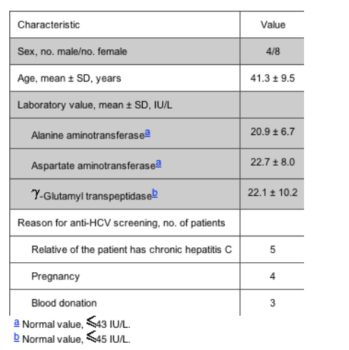
Screening for anti-HCV antibodies in these patients was done for several reasons (table 1), and, thereafter, the patients were referred to our institution, where the presence of anti-HCV antibodies was confirmed for all case patients by use of a commercial recombinant immunoblot assay (Innolia HCV Ab III; Innogenetics). In addition, all patients were serum HCV RNA negative, as was determined by a commercial test (Amplicor HCV, version 2.0 [Roche Diagnostics]; test sensitivity, 50 IU/mL; test specificity, 99%), and all had normal transaminase levels. None of these 12 patients had a clinical history of acute or chronic liver disease, and, except for 2 patients who had received a blood transfusion >25 years previously, they did not report risk factors for HCV infection. This first visit to our institution was considered to be the first day of the follow-up, which had a mean duration (±SD) of 29.2 ± 19.8 months. During the whole follow-up, the 12 anti-HCV antibody-positive patients continued to have persistently normal ALT levels and were serum HCV RNA negative, as was determined every 6 months. As was previously mentioned, a liver biopsy specimen was obtained from all these patients at a mean time (±SD) of 22.1 ± 13.6 months after the beginning of the follow-up. Table 2 presents data on transaminase levels at 3 time points during follow-up, for each of the 12 patients.
After the liver biopsy fragment was obtained, it was cut into 2 portions. The first portion was used for histological diagnosis [11], and the second portion was submerged (no later than 30 s after the liver specimen was obtained) into RNAlater (Ambion) and then was stored at -20 C until it was used for HCV RNA detection.
Serum and PBMC samples were collected from all patients on the same day when laparoscopy was performed. Transaminase levels were tested again, and anti-HCV antibody titers were determined by means of serial dilutions (1 : 100 to 1 : 100,000) of the serum samples, by use of a commercial kit (Innotest-HCV Ab IV; Innogenetics). Finally, an aliquot of serum was stored at -80 C, whereas PBMCs were stored in RNAlater (Ambion) at -20 C until they were used for HCV RNA detection.
Detection of genomic and antigenomic HCV RNA strands by quantitative, strand-specific, real-time reverse-transcription-polymerase chain reaction (RT-PCR). Total RNA was isolated from 200 L of serum, from liver specimens, and from PBMC samples, by use of the SV Total RNA Isolation kit (Promega). After precipitation, the pellet was dissolved in diethyl pyrocarbonate-treated distilled water. The total RNA concentration from the liver and PBMC samples was determined using spectrophotometry.
Quantification of the 5 noncoding region of the genomic and antigenomic HCV RNA strands was done using a strand-specific real-time RT-PCR, with use of the thermostable enzyme Tth for the synthesis of cDNA at a high temperature. Thus, for the amplification of the genomic HCV RNA strand, cDNA was generated in 20 L of reaction mixture that contained the total RNA extracted from 200 uL of serum or 0.5 ug of total RNA from liver specimens or PBMC samples, 50 pmol/L antisense primer UTRLC2 (5'-CAAGCACCCTATCAGGCAGT-3'), 1 mmol/L MnCl2, 200 umol/L each deoxynucleotide triphosphate, 1X RT buffer (Applied Biosystems), and 5 U of Tth (Applied Biosystems). After 20 min at 65 C, the RT activity was inactivated by Mn2+ chelation with 8 uL of the 10X chelating buffer (Applied Biosystems), followed by heating at 95 C for 30 min. For amplification of antigenomic HCV RNA, cDNA was synthesized under the same conditions by the addition of 50 pmol/L sense primer UTRLC1 (5'-CTTCACGCAGAAAGCGTCTA-3'). Real-time PCR was run in a LightCycler (Roche Molecular Biochemicals) with 2 L of cDNA in a final volume of 20 uL, with use of the LightCycler FastStart DNA Master SYBR Green I kit (Roche Molecular Biochemicals). The reaction mixture contained 4 mmol/L MgCl2, 0.5 umol/L primers UTRLC1 and UTRLC2, and 2 uL of SYBR Green Master mix. Amplification was performed as follows: initial denaturation and activation of enzyme were performed at 95 C for 10 min; followed by 60 cycles at 95 C for 1 s, at 60 C for 5 s, and at 72 C for 10 s; and then followed by a final step of fluorescence acquisition performed at 89 C for 5 s.
Two standard curves constructed with 10-fold dilutions of synthetic genomic and antigenomic HCV RNA (from 3.2 X 10⁸ to 0.32 copies) were used for the quantification of both HCV RNA stands. The specificity of the assay for the detection of the antigenomic HCV RNA strand was assessed by performing RT with the sense primer and serial dilutions of the synthetic genomic HCV RNA as template. Linearity of the quantification assay ranged from 3.2 to 3.2 X 10⁸ copies of genomic or antigenomic HCV RNA strand per reaction (figure 1). This assay was capable of detecting 3.2 molecules of the correct strand while unspecifically detecting 107-108 copies of the incorrect strand. The sensitivity and dynamics of each assay were not affected when total RNA extracted from HepG2 cells was added to the reaction.
|
|
| |
| |
|
|
|