| |
Bone Disease and HIV Infection
|
| |
| |
Clinical Infectious Diseases July 1, 2006;42:108-114
Valerianna Amorosa and Pablo Tebas
Division of Infectious Diseases, University of Pennsylvania School of Medicine, Philadelphia
".....Ideally, individuals at risk for fragility fracture should be selected (for screening), although the frequency of osteoporosis and osteopenia among HIV-infected individuals is so high that HIV infection might in the future be considered an indication for evaluation of BMD. Although the HIV-related risk factors for osteopenia continue to be discerned, results of longitudinal studies show that duration of HIV disease, low BMI, history of weight loss, and previous use of steroids appear to pose the major risk [4, 8, 20, 48]. In addition, we can assess other traditional risk factors that likely correlate with risk, as noted above and in table 1 [17]. Moreover, a nutritional assessment for adequate daily calcium and vitamin D intake is appropriate..... Patients with significant risk factors for fragility fracture should be screened by DEXA and considered for treatment. Rather than treating all patients with reduced BMD, patients with previous fragility fracture and significant risk factors for future fracture may be at highest priority to treat...."
The high prevalence of bone demineralization among human immunodeficiency virus (HIV)-infected patients in the current therapeutic era has been described in multiple studies, sounding the alarm that we may expect an epidemic of fragility fractures in the future. However, despite noting high overall prevalences of osteopenia and osteoporosis, recent longitudinal studies that we review here have generally not observed accelerated bone loss during antiretroviral therapy beyond the initial period after treatment initiation (note from Jules Levin: I'm not convinced studies show this; I see 1 study suggested this, the Gilead 903 study). We discuss the continued progress toward understanding the mechanisms of HIV-associated bone loss, particularly the effects of HIV infection, antiretroviral therapy, and host immune factors on bone turnover. We summarize results of clinical trials published in the past year that studied the safety and efficacy of treatment of bone loss in HIV-infected patients and provide provisional opinions about who should be considered for bone disease screening and treatment.
With the dramatic improvement in life expectancy among HIV-infected patients in the HAART era, concern has emerged regarding long-term consequences of chronic HIV infection and antiretroviral therapy (ART). The high prevalence of bone demineralization among HIV-infected patients has been described in multiple studies [112], although longitudinal studies have not generally observed accelerated bone loss beyond the initial period after ART initiation.
Given the complex metabolic complications of HIV infection and its treatment, the decreased bone mineralization seen in a large percentage of HIV-infected patients is likely the result of heterogeneous causes and interplay of host, viral, and specific antiretroviral factors. Bone is constantly undergoing remodeling in a synchronized balance between resorption and formation, which can become unregulated during HIV infection [6, 8, 13, 14].
Bone is composed of matrix and osteoid, and it is mineralized with calcium and phosphate in the form of calcium hydroxyapatite. When bone mineralization is decreased, osteopenia can occur. Eventually, osteoporosis (i.e., porous bone) can result. This is seen pathologically as the structural deterioration of bone and can lead to nontraumatic fractures. Osteomalacia (i.e., soft bone) is most commonly the result of vitamin D deficiency and occurs when intact bone matrix is not adequately mineralized.
On the basis of results of large epidemiologic studies that clearly correlated the risk of decreased bone mineral density (BMD; as measured by dual X-ray absorptiometry [DEXA]) with an increased incidence of fragility fracture [15], a World Health Organization committee provided definitions of osteoporosis and osteopenia that were based on the SD between a patient's BMD and the mean BMD at the time of peak bone mass among 30-year-old persons, adjusting for race and sex. A z score is normalized for age, as well. Osteoporosis is defined as a T score of less than -2.5 SDs. Osteopenia is defined as T score between -2.5 and -1 SDs. Multiple recent studies involving HIV-infected patients have consistently found a high prevalence of osteopenia and osteoporosis, as assessed by these criteria [112].
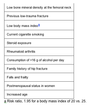
The validated risk factors recognized for fragility fracture in population studies are noted in table 1, with low-trauma fracture (i.e., fracture that occurs from a fall from standing height or less) as a strong predictor of future fracture [16, 17]. Other factors that can lead to accelerated bone loss are vitamin D deficiency, other nutritional deficiencies, low levels of calcium intake, immobilization, hypogonadism from multiple etiologies, hyperthyroidism, hyperparathyroidism, renal insufficiency, and use of certain medications, particularly anticonvulsants. Opiate use can contribute to secondary hypogonadism [18] that, in turn, could lead to increased osteopenia and osteoporosis, which is seen among active injection-heroin users [19]. Beyond these attributable risks, prolonged HIV infection is associated with decreased BMD [8, 11, 20]. No specific associations between opportunistic infections and osteoporosis have been noted, although with opportunistic infections, patients may have many risks for decreased BMD and fracture, such as low body weight, frailty, immobilization, advanced HIV infection, hypogonadism, or steroid treatment.
Table 1. Validated risk factors for fragility fracture.
For a given decrease in BMD, the fracture risk increases dramatically with age [21]. HIV-infected populations are relatively young, and an epidemic of fragility fractures has not been described among them, although reports suggest that such fractures are occurring [12, 2224]. As HIV-infected populations age, one expects an increase in the incidence of fragility fractures and the morbidity and mortality with which they are associated.
ETIOLOGY
The role of HIV infection. Early reports of the effects of HIV infection on bone metabolism did not demonstrate a dramatic increase in the prevalence of osteoporosis and osteopenia among patients with HIV infection, compared with patients without HIV infection [13, 25, 26], although small sample sizes limited the strength of their conclusions. Recently, the contribution of HIV infection to bone demineralization has been clarified further, with publication of the baseline BMD data from Gilead Study 903 [27]. This study, a randomized controlled trial of 600 ART-naive patients who received either tenofovir/lamivudine/efavirenz or stavudine/lamivudine/efavirenz during a period of 144 weeks, was the largest ever undertaken that evaluated BMD in treatment-naive individuals, and it demonstrated that HIV infection plays a significant role in the bone abnormalities seen in HIV-infected patients. The investigators found a baseline prevalence of osteopenia of 23% in the tenofovir group and 28% in the stavudine group (mean age of subjects, 36 years) [28], which is significantly higher than the national prevalence among US adults [29]. Lower baseline lumbar spine T scores correlated with lower weight, male sex, and increased age.
The role of treatment. Longitudinal studies avoid the inherent biases of descriptions from earlier cross-sectional studies of bone abnormalities in HIV-infected individuals. Several studies have now examined BMD over time in patients receiving HAART [4, 8, 20]. Nolan et al. [4] studied 54 patients for a period of 54 weeks, noting that a lower pretreatment BMI was associated with a lower baseline BMD. Neither indinavir nor nelfinavir-based regimens were associated with decreased BMD over time (indinavir therapy was associated with an annual increase of 0.31 in the z score, and there was no clinically significant change associated with nelfinavir treatment). In a longitudinal study of 12 ART-naive adults initiating ART with abacavir/lamivudine and amprenavir, Dube et al. [30] found that, at week 48 of the follow-up period, the total body BMD had significantly increased. In a study in which 93 patients receiving HAART were studied for 72 weeks, Mondy et al. [8] noted that the most significant factors associated with a low baseline BMD were low BMI, low body weight, long duration of HIV infection (>7 years since diagnosis), and smoking history. During the study period, the overall BMD increased significantly at the spine and hip and was not associated with protease inhibitor therapy [8]. During the intervals examined, these studies demonstrated a stable or slightly improved BMD for patients receiving stable ART.
It is now generally accepted that protease inhibitors do not cause osteopenia and osteoporosis. Studies in which ART was switched to nonprotease inhibitor based regimens did not affect BMD support this idea. In Adult AIDS Clinical Trials Group 5125, a total of 62 patients were randomly assigned to switch from a protease inhibitorbased regimen to a nonnucleoside reverse-transcriptase inhibitorbased regimen or to a NRTI-sparing regimen containing lopinavir/ritonavir and efavirenz [31]. After a median follow-up period of 104 weeks, there was no significant change in BMD within or between groups. Earlier, our group evaluated BMD longitudinally during a 48-week period for 18 patients who switched from protease inhibitorbased regimens to nevirapine-containing, protease inhibitorsparing regimens and found no significant changes in BMD during the study period [32].
Nonetheless, initiation of ART seems to be associated with some bone loss, the quantity of which varies with respect to treatment regimen. In Gilead Study 903 described above, during the 144 weeks, BMD of the spine decreased by 2.2% in the tenofovir arm, compared with 1.0% in the stavudine arm (P < .001), and BMD of the hip decreased by 2.8% in the tenofovir arm, compared with 2.4% in the stavudine arm (P = .064) [27]. The overall prevalence of osteoporosis and osteopenia increased slightly by the end of the study, with a prevalence of osteopenia of 28% in the tenofovir group and 27% in the stavudine group (absolute increase from normal to osteopenia, 13% vs. 8%) and a prevalence of osteoporosis of 5% in each group (absolute increase, 0 vs. <1%) [28]. This initial, modest loss of bone after initiation of therapy tends to stabilize over time, and its clinical significance and implications for regimens of intermittent ART remain unclear.
With regard to the effect that specific ARTs have on BMD, findings from Gilead Study 903 and other observational studies suggest that tenofovir may have more deleterious effects on bone than do other studied antiretrovirals, although the clinical significance of these findings is as yet unclear [20, 27]. A longitudinal study of 19 HIV-infected children demonstrated significantly decreased BMD during a 48-week course of tenofovir [33]. Tenofovir is capable of causing a Fanconi-like syndrome with renal phosphate wasting and concomitant osteomalacia, particularly when present at supratherapeutic levels for a prolonged duration, as shown in a study of rhesus macaques [34]. Although reports of Fanconi syndrome were more common with high-dose adefovir, reports of Fanconi syndrome associated with tenofovir are emerging as well [35]. There are ongoing clinical trials to better examine the effect of tenofovir on BMD and bone turnover in children.
With regard to the effects of specific protease inhibitors on BMD, Fakruddin et al. [36] demonstrated increased osteoclast differentiation peripherally in women receiving ritonavir-containing regimens, compared with women who were ART naive or receiving other ART regimens. Fakruddin and colleagues noted increased levels of markers of bone turnover (osteocalcin, bone-specific alkaline phosphatase, and urine N-telopeptide), as well as decreased BMD of the lumbar spine in ritonavir-treated patients. Also of concern and, perhaps, of greater clinical relevance, ritonavir's inhibition of cytochrome enzymatic activity could lead to osteoporosis via effects on exogenous hormone metabolism, as illustrated in a recent case series in which osteoporosis and Cushing syndrome developed in 6 patients who were receiving inhaled corticosteroids while taking a ritonavir-boosted protease inhibitor [37].
Effects on vitamin D are another mechanism by which antiretrovirals may influence bone metabolism. Calcitriol (1,25-dihydroxyvitamin D), the steroid hormone that promotes intestinal calcium absorption and regulates osteoblast function, becomes activated by cytochrome enzymes in the liver, the kidneys, and macrophages. One study noted lower levels of calcitriol in patients with advanced HIV infection, compared with patients with nonadvanced HIV infection [38], and another reported lower levels in HIV-infected patients receiving HAART, compared with control subjects [12]. Table 2 summarizes in vitro findings of the effects of protease inhibitors on vitamin D metabolism. These findings suggest that measuring the activated vitamin D levels and correcting vitamin D deficiency are important components for assessing and treating decreased BMD in HIV-infected patients.
PREVALENCE OF OSTEOPENIA AND OSTEOPOROSIS AMONG WOMEN AND AMONG CHILDREN
Bone density in HIV-infected women has been directly examined. Huang et al. [47] noted that reduced BMD in HIV-infected women with wasting was associated with reduced muscle mass. The same investigators then assessed BMD in 84 HIV-infected women and 63 HIV-uninfected women with a mean age of 42 years and a body weight within the normal range and detected a lower BMD of the lumbar spine and hip for the HIV-infected women by means of DEXA [48]. Osteopenia was present in 54% of the HIV-infected women versus 30% of the control subjects, and osteoporosis was present in 10% versus 5%. Total body fat was significantly lower for HIV-infected women than for controls, although lean body mass was similar. Among HIV-infected women, BMI, body fat and lean body mass, and lowest adult weight were positively correlated with BMD of the hip and lumbar spine. BMD in oligomenorrheic women was lower than that in eumenorrheic women, suggesting that oligomenorrhea contributes to decreased BMD in some women. Duration and class of ART were not predictors of decreased BMD.
Among 31 HIV-infected African-American and Hispanic postmenopausal, HIV-infected women, Yin et al. [49] noted a 42% prevalence of osteoporosis at the spine, compared with 23% for historical control subjects (P = .03). Of concern, the prevalence of osteoporosis at the hip was 10% among the postmenopausal, HIV-infected subjects, compared with 1% among the historical control subjects (P = .003). Instead of HIV-related risk factors, such as CD4 cell count or ART history, time since menopause onset, and weight were statistically significant predictors of BMD.
Another group with a potentially high risk for future fractures is HIV-infected children, for whom several studies have noted decreased BMD by means of DEXA [38, 5052]. Several groups have now studied HIV-infected children longitudinally for evidence of bone loss. After an earlier cross-sectional study in which the BMD for HIV-infected children receiving HAART was observed to be less than that for HAART-naive children and healthy children [38], Mora et al. [52] compared BMDs in a cohort of 32 HIV-infected children receiving HAART and healthy control subjects longitudinally during a 1-year period. They found a lower baseline BMD among HIV-infected patients, relatively similar 1-year incremental increases in BMD of the lumbar spine in both groups, and a smaller total body BMD among HIV-infected patients. Mora and colleagues also found increased markers of both bone resorption and formation in the HIV-infected group. The high rate of bone turnover did not improve longitudinally during receipt of HAART [52]. More longitudinal data are needed to ascertain the clinical significance of these findings. However, the concern is that HIV-infected children will not achieve adequate peak bone mass.
PATHOGENESIS
Determination of the pathogenesis of decreased BMD for HIV-infected patients is important to identify modifiable risk factors and innovative treatments and to appropriately select patients for screening and treatment. Bone turnover is related to bone resorption, formation, and strength. Normally, bone formation and resorption are closely linked and synchronized. Bone remodeling depends on the coupled activity of osteoblasts, which form new bone and osteoclasts (i.e., cells of monocyte or macrophage origin that degrade the bone matrix). The balance between osteoblast and osteoclast activity is a key determinant of bone mass and fracture risk. Several factors regulate osteoclast number and activity, including hormones and inflammatory cytokines via cellular signaling pathways.
Bone biopsy for histomorphometric analysis of the iliac crest is the gold standard for assessing bone turnover, but because it is an invasive procedure, biochemical markers of bone formation (e.g., levels of osteocalcin and bone-specific alkaline phosphatase) and resorption (e.g., levels of urinary deoxypyridinoline and the N-terminal and C-terminal telopeptides of type I collagen) are used to assess bone turnover in clinical studies. In some large studies comprised of women from different postmenopausal populations, increased levels of resorptive markers have been shown to correlate with fracture risk [53], and in the future, a more clearly defined role for certain markers in clinical practice will likely be established.
Several studies examining markers of bone resorption and formation have noted an uncoupling of these events in persons with advanced HIV infection. In the pre-HAART era, Serrano et al. [13] performed bone biopsies and measured markers of bone turnover in 22 HIV-infected patients and found overall reduced bone formation, with decreased numbers of osteoclasts and without clinically significantly decreased mineralization. Levels of osteocalcin, a bone formation marker, directly correlated with CD4 cell count [13]. Analyzing data for 50 women receiving 1 NRTI or no therapy, Teichman et al. [6] demonstrated a significant increase in bone resorption and a decrease in bone formation, compared with health control subjects. They also noted a positive correlation between CD4 cell count and markers of formation and a negative correlation between CD4 cell count and resorptive markers. In the HAART era, Aukrust et al. [14] examined markers of bone formation and resorption longitudinally for 16 patients after HAART initiation. Analysis of data after 24 weeks of follow-up demonstrated that, although there was no relationship between resorption and formation before therapy, there was a significant correlation between resorption and formation developed during therapy, suggesting a recoupling of resorption and bone formation and, perhaps, normalization of bone metabolism during HAART.
In the 72-week longitudinal study described above, Mondy et al. [8] found that levels of bone resorption and formation markers remained high throughout the study period, suggesting a high bone turnover state, whereas BMD slightly increased over time. Among 7 patients for whom iliac crest bone biopsies were performed, varied etiologies for decreased BMD were evident, including osteomalacia, high-turnover osteoporosis with elevated osteoblast content, inactive osteoporosis with decreased numbers of osteoblasts and osteoclasts, and osteoporosis with normal rates of remodeling and turnover. The various pathologic findings for these 7 patients suggest that bone loss is the final pathway for a diversity of causes in HIV-infected patients.
The role of cytokine activation in increasing bone resorption has been suggested, given the association of chronic cytokine activation in persons with rheumatoid arthritis and osteoporosis. There has been progress in our understanding of the role of cytokine-induced pathways in the increased bone resorption seen in some persons with advanced HIV infection. Receptor of activated NF-Kbeta ligand (RANKL) is a cytokine secreted from T cells and osteoblasts that stimulates osteoclast precursors to differentiate into osteoclasts. RANKL recruits TNF receptor-associated factor 6 to the cytoplasmic portion of the receptor of activated NF-Kbeta within osteoclasts and precursors, leading to activation of NF-Kbeta and other pathways that directly cause osteoclast differentiation and survival and bone resorption. TNF also leads to upregulation of RANKL and to induction of osteoclastogenesis [54]. Because advanced HIV disease correlates with high levels of TNF [55] and because patients with advanced HIV infection (defined as a mean CD4 cell count of 20 cells/mL) have markers of bone resorption that positively correlate with activation of TNF [14], increased bone resorption in some patients may be due to increased cytokine activation and may contribute to decreased BMD. Fakruddin et al. [36] demonstrated higher levels of RANKL in serum specimens from HIV-infected women than in serum specimens from HIV-uninfected women, regardless of ART use, suggesting a role for this cytokine pathway in HIV-associated bone loss.
In vitro data elucidating viral and antiretroviral involvement in these pathways is emerging and still often conflicting. Table 2 summarizes results of recent in vitro and animal studies. Although these findings provide insights into the mechanisms by which ART may lead to bone loss, they cannot predict clinical impact. In addition to any negative effects ART might have on bone metabolism, there are potentially positive roles associated with antagonizing the effects of chronic viremia and inflammation. This has been borne out in longitudinal studies of patients receiving various ART regimens who were not experiencing significant BMD loss over time [4, 8, 20, 28, 31, 32].
TREATMENT
Given the high prevalence of osteopenia and osteoporosis, whom should clinicians screen? Ideally, individuals at risk for fragility fracture should be selected, although the frequency of osteoporosis and osteopenia among HIV-infected individuals is so high that HIV infection might in the future be considered an indication for evaluation of BMD. Although the HIV-related risk factors for osteopenia continue to be discerned, results of longitudinal studies show that duration of HIV disease, low BMI, history of weight loss, and previous use of steroids appear to pose the major risk [4, 8, 20, 48]. In addition, we can assess other traditional risk factors that likely correlate with risk, as noted above and in table 1 [17]. Moreover, a nutritional assessment for adequate daily calcium and vitamin D intake is appropriate.
Patients with established low-trauma fractures are at highest risk for recurrent fracture and are a priority for mitigation of modifiable risk factors and treatment [16]. There are as yet no strongly compelling data from clinical trials to tailor antiretroviral therapy on the basis of BMD considerations, although tenofovir treatment may not be the first choice when there is significant concern, particularly in children [33]. The early associations seen between protease inhibitors and decreased BMD have not been consistently found in later longitudinal clinical trials [4, 31, 32].
Note from Jules Levin; recent reports found in I think a small number of patients Fosomax associated with loss of bone in the jaw.
A number of studies have now confirmed the safety and efficacy of bisphosphonates in the treatment of osteopenia and osteoporosis in persons with HIV infection [5658]. Mondy et al. [56] conducted a 48-week randomized trial involving 31 HIV-infected patients with osteopenia or osteoporosis (T score, less than -1) receiving HAART in which treatment with alendronate (70 mg each week) together with calcium (1000 mg of calcium carbonate daily) and vitamin D (400 IU daily) was compared with calcium/vitamin D therapy alone. Mondy and colleagues noted a 5.2% increase (95% CI, 1.3%6.4%) in the BMD of the lumber spine for the alendronate-treated group, compared with a 1.3% increase (95% CI, -2.4% to 4.0%) for the vitamin D/calcium alone group. Negredo et al. [57] observed 25 HIV-infected patients receiving alendronate for 96 weeks and noted a reduction in the prevalence of osteoporosis and improvement in the trochanteric BMD in the alendronate group. Guaraldi et al. [58] conducted a 42-patient randomized trial during a 52-week period and noted increases in BMD of the lumber spine and a deceleration of bone loss at the hip in the alendronate group. A larger randomized, multicenter trial (Adult AIDS Clinical Trials Group 5163) examining alendronate continues, with results to be made available in 2006.
Treatment with raloxifene, a selective estrogen-receptor modulator used in postmenopausal women, should be considered only with caution, given the demonstrated cytochrome P450 inhibition. With their potential for drug interactions, the use of raloxifene and other hormonal therapies will require further study in HIV-infected patients. Fairfield et al. [59] noted that testosterone therapy in eugonadal men with concomitant wasting increased BMD, although at this point, hypogonadism is the most appropriate indication for its use. The use of growth hormonereleasing hormone for patients with concomitant fat redistribution requires further study but may prove to be a promising option in the future [60].
CONCLUSION
There are several questions regarding screening and treatment of low BMD in HIV-infected individuals that still lack definitive answers. Patients with significant risk factors for fragility fracture should be screened by DEXA and considered for treatment. Rather than treating all patients with reduced BMD, patients with previous fragility fracture and significant risk factors for future fracture may be at highest priority to treat. More longitudinal data on treatment duration and outcomes and the natural history of osteopenia and osteoporosis in HIV-infected patients will aid in establishing appropriate thresholds for pharmacologic interventions.
References
1. Tebas P, Powderly WG, Claxton S, et al. Accelerated bone mineral loss in HIV-infected patients receiving potent antiretroviral therapy. AIDS 2000; 14:F637. First citation in
2. Carr A, Miller J, Eisman JA, Cooper DA. Osteopenia in HIV-infected men: association with asymptomatic lactic acidemia and lower weight pre-antiretroviral therapy. AIDS 2001; 15:7039. First citation in
3. Moore AL, Vashisht A, Sabin CA, et al. Reduced bone mineral density in HIV-positive individuals. AIDS 2001; 15:17313. First citation in
4. Nolan D, Upton R, McKinnon E, et al. Stable or increasing bone mineral density in HIV-infected patients treated with nelfinavir or indinavir. AIDS 2001; 15:127580. First citation in
5. Huang JS, Rietschel P, Hadigan CM, Rosenthal DI, Grinspoon S. Increased abdominal visceral fat is associated with reduced bone density in HIV-infected men with lipodystrophy. AIDS 2001; 15:97582. First citation in
6. Teichmann J, Stephan E, Lange U, et al. Osteopenia in HIV-infected women prior to highly active antiretroviral therapy. J Infect 2003; 46:2217. First citation in
7. Landonio S, Quirino T, Bonfanti P, et al. Osteopenia and osteoporosis in HIV+ patients, untreated or receiving HAART. Biomed Pharmacother 2004; 58:5058. First citation in
8. Mondy K, Yarasheski K, Powderly WG, et al. Longitudinal evolution of bone mineral density and bone markers in HIV-infected individuals. Clin Infect Dis 2003; 36:48290. First citation in
| Full Text |
9. Brown TT, Ruppe MD, Kassner R, et al. Reduced bone mineral density in human immunodeficiency virusinfected patients and its association with increased central adiposity and postload hyperglycemia. J Clin Endocrinol Metab 2004; 89:12006. First citation in
10. Amiel C, Ostertag A, Slama L, et al. BMD is reduced in HIV-infected men irrespective of treatment. J Bone Miner Res 2004; 19:4029. First citation in
|
11. Bruera D, Luna N, David DO, Bergoglio LM, Zamudio J. Decreased bone mineral density in HIV-infected patients is independent of antiretroviral therapy. AIDS 2003; 17:191723. First citation in
12. Madeddu G, Spanu A, Solinas P, et al. Bone mass loss and vitamin D metabolism impairment in HIV patients receiving highly active antiretroviral therapy. Q J Nucl Med Mol Imaging 2004; 48:3948. First citation in
|
13. Serrano S, Marinoso M, Soriano J, et al. Bone remodelling in human immunodeficiency virus-linfected patients: a histomorphometric study. Bone 1995; 16:18591. First citation in
14. Aukrust P, Haug C, Ueland T, et al. Decreased bone formative and enhanced resorptive markers in human immunodeficiency virus infection: indication of normalization of the bone-remodeling process during highly active antiretroviral therapy. J Clin Endocrinol Metab 1999; 84:14550. First citation in
15. Cummings SR, Black DM, Nevitt MC, et al. Bone density at various sites for prediction of hip fractures. The Study of Osteoporotic Fractures Research Group. Lancet 1993; 341:725. First citation in
16. Ross PD, Davis JW, Epstein RS, Wasnich RD. Preexisting fractures and bone mass predict vertebral fracture incidence in women. Ann Intern Med 1991; 114:91923. First citation in
|
17. Kanis JA, Borgstrom F, De Laet C, et al. Assessment of fracture risk. Osteoporos Int 2005; 16:5819. First citation in
18. Cooper OB, Brown TT, Dobs AS. Opiate drug use: a potential contributor to the endocrine and metabolic complications in human immunodeficiency virus disease. Clin Infect Dis 2003; 37(Suppl 2):S1326. First citation in
| Full Text |
19. Pedrazzoni M, Vescovi PP, Maninetti L, et al. Effects of chronic heroine abuse on bone and mineral metabolism. Acta Endocrinol (Copenh) 1993; 129:425. First citation in
|
20. Jacobson D, Huang J, Shevitz A, Knox T, Gorbach S, Spiegelman D. Duration of ART and change in bone mineral density over time [abstract 825]. In: Program and abstracts of the 12th Conference on Retroviruses and Opportunistic Infections (Boston). Alexandria, VA: Foundation for Retrovirology and Human Health, 2005. First citation in
21. Kanis JA, Johnell O, Oden A, Dawson A, De Laet C, Jonsson B. T, Ten year probabilities of osteoporotic fractures according to BMD and diagnostic thresholds. Osteoporos Int 2001; 12:98995. First citation in
22. Martin K, Lawson-Ayayi S, Miremont-Salame G, et al. Symptomatic bone disorders in HIV-infected patients: incidence in the Aquitaine cohort (19992002). Groupe d'Epidemiologie Clinique du SIDA en Aquitaine. HIV Med 2004; 5:4216. First citation in
23. McComsey G, Huang J, Woolley I, et al. Fragility fractures in HIV-infected subjects, an area for improvement [abstract 743]. In: Program and abstracts of the 11th Conference on Retroviruses and Opportunistic Infections (San Francisco). Alexandria, VA: Foundation for Retrovirology and Human Health, 2004. First citation in
24. Stephens EA, Das R, Madge S, et al. Symptomatic osteoporosis in two young HIV-positive African women. AIDS 1999; 13:26056. First citation in
25. Hernandez Quero J, Ortego Centeno N, Munoz Torre M, Martinez Perez MA, Torres-Puchol JM. Alterations in bone turnover in HIV-positive patients. Infection 1993; 21:2202. First citation in
26. Paton NIJ, Macallan D, Griffin GE, Pazianas M. Bone mineral density in patients with human immunodeficiency virus infection. Calcif Tissue Int 1997; 61:302. First citation in
27. Gallant JE, Staszewski S, Pozniak AL, et al. Efficacy and safety of tenofovir DF vs. stavudine in combination therapy in antiretroviral-naive patients: a 3-year randomized trial. 903 Study Group. JAMA 2004; 292:191201. First citation in
28. Powderly W, Cohen C, Gallant J, Lu B, Enejosa J, Cheng A. Similar incidence of osteopenia and osteoporosis in ART-naive patients treated with tenofovir DF or stavudine in combination with lamivudine and efavirenz over 144 weeks [abstract 823]. 903 Study Group. In: Program and abstracts of the 12th Conference on Retroviruses and Opportunistic Infections (Boston). Alexandria, VA: Foundation for Retrovirology and Human Health, 2005. First citation in
29. Looker AC, Wahner H, Dunn WL, et al. Updated data on proximal femur bone mineral levels of US adults. Osteoporo Int 1998; 8:46889. First citation in
30. Dube MP, Qian D, Edmondson-Melancon H, et al. Prospective, intensive study of metabolic changes associated with 48 weeks of amprenavir-based antiretroviral therapy. Clin Infect Dis 2002; 35:47581. First citation in
| Full Text |
31. Tebas P, Zhang J, Yarasheski K, et al. Switch to a protease inhibitorcontaining/nucleoside reverse-transcriptase inhibitorsparing regimen increases appendicular fat and serum lipid levels without affecting glucose metabolism or bone mineral density: the results of a prospective randomized trial, ACTG 5125s [abstract 40]. Adult AIDS Clinical Trials Group. In: Program and abstracts of the 12th Conference on Retroviruses and Opportunistic Infections (Boston). Alexandria, VA: Foundation for Retrovirology and Human Health, 2005. First citation in
32. Tebas P, Yarasheski K, Henry K, et al. Evaluation of the virological and metabolic effects of switching protease inhibitor combination antiretroviral therapy to nevirapine-based therapy for the treatment of HIV infection. AIDS Res Hum Retroviruses 2004; 20:58994. First citation in
33. Hazra R, Gafni R, Maldarelli F, et al. Safety, tolerability, and clinical responses to tenofovir DF in combination with other antiretrovirals in heavily treatmentexperienced HIV-infected children: data through 48 weeks [abstract 928]. In: Program and abstracts of the 11th Conference on Retroviruses and Opportunistic Infections (San Francisco). Alexandria, VA: Foundation for Retrovirology and Human Health, 2004. First citation in
34. Van Rompay KK, Brignolo LL, Meyer DJ, et al. Biological effects of short-term or prolonged administration of 9-[2(phosphonomethoxy)propyl] adenine (tenofovir) to newborn and infant rhesus macaques. Antimicrob Agents Chemother 2004; 48:146987. First citation in
35. Earle KE, Seneviratne T, Shaker J, Shoback D. Fanconi's syndrome in HIV+ adults: report of three cases and literature review. J Bone Miner Res 2004; 19:71421. First citation in
|
36. Fakruddin M, Yin M, Laurence J. Pathophysiologic correlates of RANKL deregulation in HIV infection and its therapy [abstract 822]. In: Program and abstracts of the 12th Conference on Retroviruses and Opportunistic Infections (Boston). Alexandria, VA: Foundation for Retrovirology and Human Health, 2005. First citation in
37. Samaras K, Pett S, Gowers A, McMurchie M, Cooper DA. Iatrogenic Cushing's syndrome with osteoporosis and secondary adrenal failure in HIV-infected patients receiving inhaled corticosteroids and ritonavir-boosted protease inhibitors: six cases. J Clin Endocrinol Metab 2005; 90:43948. First citation in
38. Mora S, Sala N, Bricalli D, Zuin G, Chiumello G, Vigano A. Bone mineral loss through increased bone turnover in HIV-infected children treated with highly active antiretroviral therapy. AIDS 2001; 15:18239. First citation in
39. Fakruddin JM, Laurence J. HIV envelope gp120-mediated regulation of osteoclastogenesis via receptor activator of nuclear factor kappa B ligand (RANKL) secretion and its modulation by certain HIV protease inhibitors through interferon-gamma/RANKL cross-talk. J Biol Chem 2003; 278:48,2518. First citation in
|
40. Fakruddin JM, Laurence J. HIV-1 Vpr enhances production of receptor of activated NF-kappaB ligand (RANKL) via potentiation of glucocorticoid receptor activity. Arch Virol 2005; 150:6778. First citation in
41. Wang MW, Wei S, Teitelbaum SL, Ross FP. The HIV protease inhibitor indinavir uniquely inhibits bone formation (abstract). J Bone Miner Res 2001; 16(Suppl):S372. First citation in
42. Wang MWH, Teitelbaum SJ, Tebas P, Powderly WG, Ross FP. Indinavir administration leads to bone loss in mice [abstract 713]. In: Program and abstracts of the 9th Conference on Retroviruses and Opportunistic Infections (Seattle). Alexandria, VA: Foundation for Retrovirology and Human Health, 2002. First citation in
43. Wang MW, Wei S, Faccio R, et al. The HIV protease inhibitor ritonavir blocks osteoclastogenesis and function by impairing RANKL-induced signaling. J Clin Invest 2004; 114:20613. First citation in
44. Jain RG, Lenhard J. Select HIV protease inhibitors alter bone and fat metabolism ex vivo. J Biol Chem 2002; 277:1924750. First citation in
45. Pan G, Wu X, McKenna MA, Feng X, Nagy TR, McDonald JM. AZT enhances osteoclastogenesis and bone loss. AIDS Res Hum Retroviruses 2004; 20:60820. First citation in
46. Cozzolino M, Vidal M, Arcidiacono MV, Tebas P, Yarasheski KE, Dusso AS. HIV-protease inhibitors impair vitamin D bioactivation to 1,25-dihydroxyvitamin D. AIDS 2003; 17:51320. First citation in
47. Huang JS, Wilkie S, Sullivan MP, Grinspoon S. Reduced bone density in androgen-deficient women with acquired immune deficiency syndrome wasting. J Clin Endocrinol Metab 2001; 86:35339. First citation in
48. Dolan SE, Huang J, Killilea KM, Sullivan MP, Aliabadi N, Grinspoon S. Reduced bone density in HIV-infected women. AIDS 2004; 18:47583. First citation in
49. Yin M, Dobkin J, Brudney K, et al. Bone mass and mineral metabolism in HIV+ postmenopausal women. Osteoporos Int 2005; 16:134552. First citation in
50. Arpadi SM, Horlick M, Thornton J, Cuff PA, Wang J, Kotler DP. Bone mineral content is lower in prepubertal HIV-infected children. J Acquir Immune Defic Syndr 2002; 29:4504. First citation in
|
51. Rojo Conejo P, Ramos Amador JT, Garcia Pinar L, et al. Decreased bone mineral density in HIV-infected children receiving highly active antiretroviral therapy [in Spanish]. An Pediatr (Barc) 2004; 60:24953. First citation in
|
52. Mora S, Zamproni I, Beccio S, Bianchi R, Giacomet V, Vigano A. Longitudinal changes of bone mineral density and metabolism in antiretroviral-treated human immunodeficiency virusinfected children. J Clin Endocrinol Metab 2004; 89:248. First citation in
53. Seibel M. Biochemical markers of bone remodeling. Endocrinol Metab Clin North Am 2003; 32:83113. First citation in
54. Wei S, Kitaura H, Zhou P, Ross FP, Teitelbaum SL. IL-1 mediates TNF-induced osteoclastogenesis. J Clin Invest 2005; 115:28290. First citation in
55. Hittinger G, Poggi C, Delbeke E, Profizi N, Lafeuillade A. Correlation between plasma levels of cytokines and HIV-1 RNA copy number in HIV-infected patients. Infection 1998; 26:1003. First citation in
|
56. Mondy K, Powderly W, Claxton SA, et al. Alendronate, vitamin D, and calcium for the treatment of osteopenia/osteoporosis associated with HIV infection. J Acquir Immune Defic Syndr 2005; 38:42631. First citation in
57. Negredo E, Martinez-Lopez E, Paredes R, et al. Reversal of HIV-1associated osteoporosis with once-weekly alendronate. AIDS 2005; 19:3435. First citation in
|
58. Guaraldi G, Orlando G, Madeddu G, et al. Alendronate reduces bone resorption in HIV-associated osteopenia/osteoporosis. HIV Clin Trials 2004; 5:26977. First citation in
59. Fairfield WP, Treat M, Rosenthal DI, et al. Effects of testosterone and exercise on muscle leanness in eugonadal men with AIDS wasting. J Appl Physiol 2001; 90:216671. First citation in
|
60. Koutkia P, Canavan B, Breau J, Grinspoon S. Effects of growth hormonereleasing hormone on bone turnover in HIV-infected men with fat accumulation. J Clin Endocrinol Metab 2005; 90:215460. First citation in
|
|
| |
| |
|
|
|