 |
 |
 |
| |
Brain tCho/Cr is Elevated in Acute HIV within the First Month of Infection: 'brain inflammation in acute infection/immediate HAART may normalize or improve inflammation or perhaps prevent damage'
|
| |
| |
Reported by Jules Levin
CROI 2012
Napapon Sailasuta 1, Jintanat Ananworanich 2,3,4,5, Thep Chalermchai 2, Victor DeGrutolla 6, Sukalaya Lerdlum 3, Mantana Pothisri 3,
Somprartthana Rattanamanee 2, Edgar Busovaca 7, Serena Spudich 8, Victor Valcour 2,7, 9 * on behalf of the SEARCH 010/RV 254 protocol teams
1Huntington Medical Research Institutes, Pasadena, CA, USA. 2SEARCH-Thailand, Bangkok, Thailand. 3Faculty of Medicine, Chulalongkorn University, Bangkok, Thailand. 4United States Armed Force Research Institute of Medical Science, Bangkok, Thailand. 5HIV-NAT, The Thai Red Cross AIDS research centre, Bangkok, Thailand. 6Department of Epidemiology and Biostatistics, Harvard school of Public Health, Boston, MA, USA. 7Memory and Aging Center, Department of Neurology, UCSF, San Francisco, CA, USA. 8Department of Neurology, Yale University School of Medicine, New Haven, Ct, USA. 9Division of Geriatric Medicine, UCSF, San Francisco, CA,USA

The patients in this cohort have been infected for less than a month. This is not really early (primary) infection but super early acute infection, meaning there is likely not enough time for the brain to have substantial injury. The marker used was Choline. This probably means inflammation
(cellular infiltration) without neuronal injury (normal NAA) or microglial
involvement (normal MI, a marker for the cells in the brain that likely
are activated in chronic HIV). All were treated with HAART immediately
upon identification and the choline normalized. This likely means that
cellular infiltration is reduced or stopped.
So, this appears to be evidence for the brain (not just the CSF)
to be abnormal within weeks of HIV infection by alterations in choline
within brain tissue during the first month after infection, and this
appears to be controlled with immediate institution of HAART. Whether
this has a long-term impact on preservation of brain in not known (authors are
trying to figure this out). Unfortunately, even if it does, it is not
terribly practical to apply, since it is very hard to identify HIV so
early on a public health basis, but it does help to understand the
timing of brain involvement and recognize that the brain is a common early
target.

ABSTRACT
Background: Non-invasive imaging techniques can reveal changes in central nervous system (CNS) chemistry suggestive of inflammation in HIV. These changes have been described during primary HIV infection in humans and acute SIV infection in macaques; however, data are lacking among HIV+ individuals during acute HIV (AHI) prior to seroconversion. This study aimed to define CNS immune response and involvement in AHI using Proton Magnetic Resonance Spectroscopy (1HMRS).
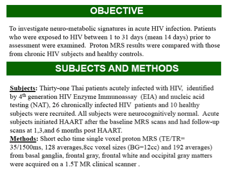
Methods: We completed four-voxel MRS among 31Thai subjects with AHI
(Fiebig I-IV) compared to cognitively normal chronic HIV-infected (CHI)
subjects who were naïve to combination antiretroviral therapy (cART) and ten HIV-negative Thai controls. MRS voxels were placed in the basal ganglia (BG), frontal grey matter (FGM), frontal white matter (FWM), and occipital grey matter (OGM) to measure myoinositol (mI), choline (tCho), N-acetyl aspartate (NAA), and glutamate/glutamine (Glx) standardized as a ratio to creatine (Cr).
Results: We evaluated during Fiebig (F) stages: F-I (10); F-II (3); F-III (15);
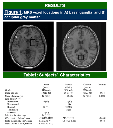
or F-IV (3). The estimated duration of infection ranged from 1-32 days, with a median of 13 days. Mean (range) age was 30 (19-46) and 26 were male. CHI subjects were mean (range) age 34 (25-48) with CD4 count of 202 (19-553) and log10 plasma HIV RNA of 4.82 (3.22-5.88). 52% were female. Control subjects were mean (range) age of 35.5 (27-45) years and 40% were female.
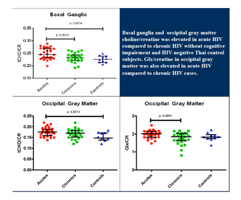
Omnibus testing identified a pattern of difference among the 3 groups in the basal ganglia (p=0.002). Adjusting for age, MRS NAA/Cr and tCho/Cr were
elevated in AHI compared to CHI (p=0.0051 and 0.0023, respectively) and
tCho/Cr was elevated in AHI vs. CO (p=0.0014). Omnibus testing also
identified differences among groups at OGM (0.0347) with elevated tCho/Cr
in AHI compared to CO (p=0.0013) and elevated Glx/Cr in AHI vs. CHI
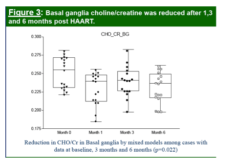
(p=0.0041). Among AHI subjects with follow-up (n=17) at six months post
HAART, BG tCho/Cr was reduced (p=0.022) and did not differ from CO
(p=0.385)*. (*last sentence updated from initial abstract to represent complete case analysis - only cases with all 3 visits included - results not changed appreciably).
Conclusions: CNS abnormalities are identified during AHI when compared to healthy control subjects and cognitively normal CHI subjects. These data provide evidence that neuroinflammation or glial dysfunction occurs in the earliest stages after exposure to HIV and are normalized by HAART.


|
| |
|
 |
 |
|
|