 |
 |
 |
| |
HIV is an Independent Predictor of Lower Bone Mineral Density in HIV-positive Subjects Compared to HIV-negative Subjects
|
| |
| |
Reported by Jules Levin
CROI 2013 Atlanta
AG Cotter,1,2 CA Sabin,3 S Simelane,1 A Macken,1 B Rogers,1 E Kavanagh,2
JJ Brady,2 PWG Mallon1,2 on behalf of the HIV UPBEAT Study Group
1HIV Molecular Research Group, School of Medicine and Medical Science, University College Dublin, Dublin, Ireland,
2Mater Misericordiae University Hospital, Dublin, Ireland
3Research Department of Infection and Population Health, University College London Medical School, London
IAS Report of further analysis from same study:
IAS: Number of regimens rather than cumulative exposure to antiretrovirals associated with lower bone mineral density in HIV-positive subjects - (07/22/13)
Excerpt from CROI Report by Todd Brown MD for NATAP - "Update from 20th CROI: Bones, Vitamin D, Frailty"
http://www.natap.org/2013/CROI/croi_145.htm
"Osteoporosis: There have been multiple studies that compare bone mineral density (BMD) in HIV+ and HIV- individuals and have generally shown lower BMD in HIV-infected persons. One of the limitations of the some of the previous studies has been the HIV-uninfected control groups, which may differ in other ways from the HIV-infected groups and may introduce bias in the comparison. To overcome this limitation, the HIV UPBEAT (Understanding the Pathology of Bone Disease in HIV-infected Subjects) recruited 210 HIV-infected subjects and 264 HIV-uninfected controls from similar demographic backgrounds (Poster #817). This Irish cohort is primarily young, male (59% HIV+ vs 44% HIV-), and Caucasian (HIV+ 61% vs HIV+ 75%). Importantly, body mass index was similar by HIV-serostatus (26 vs 27 kg/m2). BMD was found to be lower in the HIV+ group at the lumbar spine, total hip and femoral neck after adjustment for demographic factors and BMI. Low BMD (Z-score < -2) was about 2 fold higher in the HIV+ group across the sites. BMD was not related to vitamin D levels or PTH. However, alkaline phosphatase, whose skeletal derived fraction is a measure of bone turnover, was higher in HIV+ subjects and partially, but not completely, explained the difference in BMD by serostatus. This study confirms previous findings regarding low BMD in HIV+ populations. We will undoubtedly be seeing more data coming out of this rich cohort regarding the effect of antiretroviral therapy, chronic inflammation/immune activation, and other factors which may help to explain the differences by HIV-status."
ABSTRACT
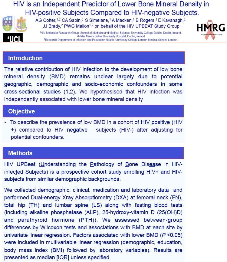
Background: The relative contribution of HIV infection to the development of low bone mineral density
(BMD) remains unclear due to a lack of adequately controlled prospective studies. We determined the
relative contribution of HIV infection to low BMD after adjustment for demographic, socio-economic and
laboratory variables.
Methods: A prospective cohort study enrolled HIV positive (HIV+) and negative (HIV-) subjects from
similar demographic backgrounds; demographic, clinical, medication and laboratory data were
collected. Dual xray absorptiometry (DXA) at femoral neck (FN), total hip (TH) and lumbar spine (LS)
and fasting blood tests (including alkaline phosphatase (ALP), 25-hydroxy-vitamin D (25(OH)D) and
parathyroid hormone (PTH)) were performed. We assessed between-group differences by Wilcoxon
tests and associations with BMD at each site by multivariable linear regression. Results are presented
as are median [IQR] unless specified.
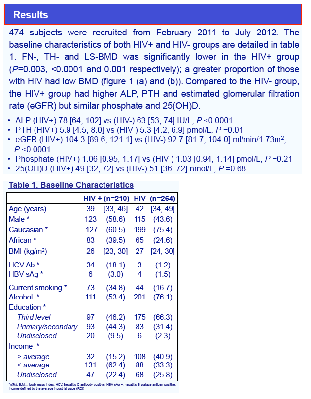
Results: 474 subjects were recruited from February 2011 to July 2012. The HIV+ group (N=210) was
58% male, 40% African, aged 39[33, 46] years; HIV acquisition risk was heterosexual sex (46.9%),
homosexual sex (25.4%), intravenous drug use (18.7%). The HIV- group (N=264) was 43% male, 25%
African and aged 42[34, 49] years. Compared to the HIV- group, the HIV+ group had higher ALP (78[64,
102] vs. 63 [53, 74]IU/L) and PTH (5.9 [4.5, 8.0] vs. 5.3 [4.2, 6.9]pmol/L, P=0.01) but similar phosphate
(1.06 [0.95, 1.17] vs. 1.03 [0.94, 1.14]mmol/L, P=0.21) and 25(OH)D (49 [32, 72] vs. 51 [36, 72]nmol/L,
P=0.68). BMD measured at FN, TH and LS was significantly lower in the HIV+ group (P=0.003, <0.0001
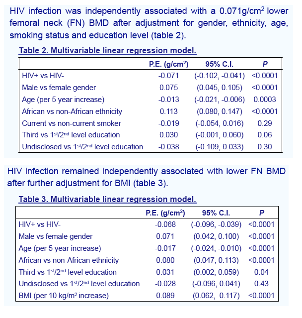
and 0.001 respectively). HIV infection was independently associated with a 0.068g/cm2 lower FN BMD
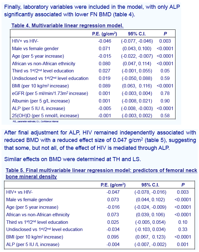
after adjustment for gender, ethnicity, age, smoking status, education and body mass index (BMI). After
further adjustment for ALP, HIV remained independently associated with reduced BMD with a reduced
effect size, 0.047 g/cm2 (table 1), suggesting that some, but not all, of the effect of HIV is mediated
through ALP. Similar effects on BMD were determined at TH and LS.
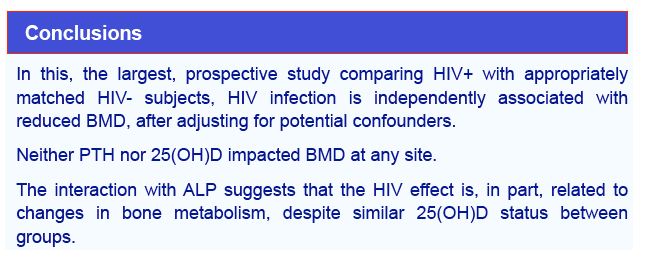
Conclusion: In this, the largest, prospective study comparing HIV+ with appropriately matched HIVsubjects,
HIV infection is independently associated with reduced BMD, after adjusting for potential
confounders. The interaction with ALP suggests that the HIV effect is, in part, related to changes in bone
metabolism, despite similar 25(OH)D status between groups.
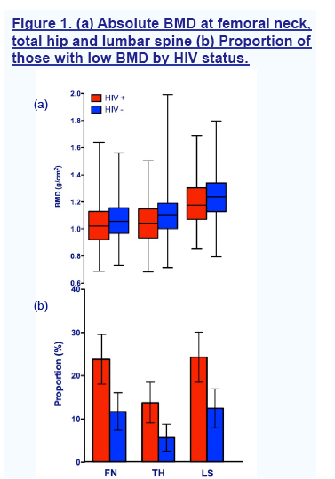
(a) Boxplots represent minimum, 25th centile, median, 75th centile and maximum BMD at each site. All between-group P <0.01. (b)
Low BMD defined as Z-score <-2.0 for those <40 years or T-score <-1.0 for those >40 years. Error bars represent 95% confidence
intervals for proportions. All between-group P <0.01. FN, femoral neck; TH, total hip; LS, lumbar spine.

|
| |
|
 |
 |
|
|