| |
Clinical Predictors of Liver Fibrosis Presence and Progression in HIV-Associated NAFLD
|
| |
| |
Download the PDF here
09 April 2020 Clinical Infectious Diseases - Lindsay T. Fourman *1, Takara L. Stanley *1, Isabel Zheng 1, Chelsea S. Pan 1, Meghan N. Feldpausch 1, Julia Purdy 2, Julia Aepfelbacher 2, Colleen Buckless 3, Andrew Tsao 3, Kathleen E. Corey 4, Raymond T. Chung 4, Martin Torriani 3,
David E. Kleiner 5, Colleen M. Hadigan 2, Steven K. Grinspoon 1
1 Metabolism Unit, Massachusetts General Hospital and Harvard Medical School, Boston, MA, USA
2 National Institute of Allergy and Infectious Diseases, National Institutes of Health, Bethesda, MD, USA
3 Department of Radiology, Massachusetts General Hospital and Harvard Medical School, Boston, MA, USA
4 Liver Center, Gastroenterology Division, Massachusetts General Hospital and Harvard Medical School, Boston, MA, USA
5 Laboratory of Pathology, Center for Cancer Research, National Cancer Institute, Bethesda, MD, USA
CROI: Clinical Predictors of Liver Fibrosis Presence and Progression in HIV-Associated Nonalcoholic Fatty Liver Disease (03/23/20)
Abstract
Background
Nonalcoholic fatty disease (NAFLD) affects over one-third of people living with HIV. Nonetheless, the natural history of HIV-associated NAFLD is poorly understood, including which patients are most likely to have a progressive disease course.
Methods
We leveraged a randomized trial of the growth hormone-releasing hormone analogue tesamorelin to treat NAFLD in HIV. Sixty-one participants with HIV-associated NAFLD were randomized to tesamorelin or placebo for 12 months. Participants underwent liver biopsy at baseline and 12 months with histologic evaluation performed by an expert pathologist blinded to treatment.
Results
In all participants with baseline biopsies (n=58), 43% had hepatic fibrosis. Individuals with fibrosis had higher NAFLD Activity Score (NAS) (3.6±2.0 vs. 2.0±0.8, P<0.0001) and visceral fat content (284±91 cm2 vs. 212±95 cm2, P=0.005), but no difference in hepatic fat or BMI. Among placebo-treated participants with paired biopsies (n=24), 38% had hepatic fibrosis progression over 12 months. For each 25 cm2 higher visceral fat at baseline, the odds of fibrosis progression increased by 37% (OR 1.37, 95% CI 1.03, 2.07). There was no difference in baseline NAS score between fibrosis progressors and non-progressors, though NAS score rose over time in the progressor group (1.1±0.8 vs. -0.5±0.6, P<0.0001).
Conclusions
In this longitudinal study of HIV-associated NAFLD, high rates of hepatic fibrosis and progression were observed. Visceral adiposity was identified as a novel clinical predictor of worsening fibrosis. In contrast, baseline histologic characteristics were not found to relate to fibrosis changes over time. Further studies are needed to identify additional biomarkers of accelerated disease.

Introduction
In an era of rising rates of obesity and hepatitis C virus (HCV) cure, nonalcoholic fatty liver disease (NAFLD) has become a leading cause of liver disease among people living with HIV (PLWH) [1]. Over one-third of individuals are affected with risk factors that include elevated BMI, metabolic comorbidities, and high CD4+ T cell count [1]. The spectrum of NAFLD is broad, ranging from simple steatosis to steatohepatitis (NASH) to fibrosis. Importantly, among patients with NAFLD in the general population, the severity of fibrosis is the strongest predictor of all-cause and liver-specific mortality [2, 3]. Thus, understanding the clinical predictors of fibrosis presence and progression in HIV is imperative so that individuals with the most aggressive hepatic disease can be appropriately monitored and targeted for intervention.
Individuals with HIV/HCV coinfection have been shown to have faster progression of fibrosis as well as a higher frequency of hepatic decompensation compared to HCV-monoinfected patients [4, 5]. While these findings raise concern that the course of NAFLD in HIV may also be accelerated, its natural history among this patient population has not been previously well-defined. In this regard, studies focused on hepatic fibrosis in the setting of HIV monoinfection have most often examined a heterogeneous sample not specifically selected for NAFLD [6-9]. Furthermore, these analyses have only rarely included long-term follow-up or utilized liver biopsies that would allow for comprehensive assessment of other histologic changes [10]. While these previous studies have consistently demonstrated high rates of hepatic fibrosis in association with metabolic risk factors [6-10], the subset of patients with HIV-associated NAFLD at highest risk for disease-related morbidity remains elusive.
In the current analysis, we leveraged phenotypic data including liver biopsy samples from a recent randomized placebo-controlled trial to characterize the longitudinal course of NAFLD in HIV. This previous study demonstrated that a strategy to reduce visceral fat prevented fibrosis progression among individuals with HIV-associated NAFLD [11]. In this current analysis, we investigated for the first time the relationship of visceral fat and other clinical characteristics with liver fibrosis, particularly with respect to the natural history of fibrosis progression among placebo-treated participants undergoing serial liver biopsies. Given the high frequency of NAFLD in HIV, accurately predicting which patients will have the most severe course of disease is critically needed to optimize screening, prevention, and treatment strategies for this population.
Results
Participant Characteristics
A total of 58 participants with HIV-associated NAFLD had liver biopsy specimens available at baseline. Clinical characteristics of the overall sample were summarized in Supplementary Table 1. Study subjects (53 ± 7 years, 81% male) had long-standing HIV infection (16 ± 9 years) that was well controlled. All participants were on stable ART with 62% receiving an integrase inhibitor-based regimen. Baseline liver fat content was 13.8% 8.6%, whereas NAS score was 2.7 ± 1.6. A total of 59% of participants were obese. BMI was strongly correlated with SAT (r = 0.86, P < 0.0001), and more weakly associated with VAT (r = 0.29, P = 0.02) (Supplementary Figure 1).
A total of 24 participants with HIV-associated NAFLD randomized to placebo had paired liver biopsy specimens available at baseline and 12 months. Clinical characteristics in this subset were comparable to the overall study group (Supplementary Table 1).
Clinical Correlates of Baseline Hepatic Fibrosis (Overall Sample)
Among our overall sample with HIV-associated NAFLD, 43% had evidence of hepatic fibrosis at baseline with the following distribution by stage: stage 1, 36%; stage 2, 40%; stage 3, 24% (Figure 1A). Clinical correlates of baseline hepatic fibrosis are shown in Table 1. Individuals with and without fibrosis were of comparable age and sex. While fibrosis tended to be more common among white subjects, racial differences between groups were not statistically significant. In contrast, there was no association of fibrosis with CD4+ T cell count, HIV viral load, C-reactive protein (CRP), or interleukin-6 (IL-6).
With regard to hepatic and metabolic indices, individuals with hepatic fibrosis had higher NAS score (3.6 ± 2.0 vs. 2.0 ± 0.8, P < 0.0001) with elevations in both lobular inflammation (1.5 ± 0.8 vs. 0.9 ± 0.2, P = 0.0004) and hepatocellular ballooning (0.7 ± 0.8 vs. 0.0 ± 0.2, P < 0.0001). In contrast, liver fat content was not found to differ between groups. Fibrosis also was positively associated with the non-invasive parameters alanine aminotransferase (ALT, 41 ± 30 U/L vs. 23 ± 8 U/L, P = 0.002), aspartate aminotransferase (AST, 44 ± 27 U/L vs. 23 ± 10 U/L, P = 0.0003), and FIB-4 (1.88 ± 0.98 vs. 1.12 ± 0.44, P = 0.0003). Notably, while VAT was higher among individuals with fibrosis (284 ± 91 cm2 vs. 212 ± 95 cm2, P = 0.005), there were no differences between groups in BMI, waist circumference, or SAT (Figure 2).
Clinical Predictors of Hepatic Fibrosis Progression (Placebo Group)
Over 12 months, fibrosis progressed in 38% (n = 9) of placebo-treated participants with HIV-associated NAFLD. Meanwhile, 50% (n = 12) of subjects had no change in fibrosis, whereas 13% (n = 3) experienced fibrosis regression (Figure 1B). Among all placebo-treated participants, the mean rate of fibrosis progression was 0.2 ± 0.8 stages per year. A total of 56% (n = 5) of participants with fibrosis progression had no evidence of fibrosis at baseline.
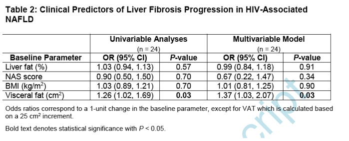
Baseline VAT was higher among those with fibrosis progression than without progression (306 ± 119 cm2 vs. 212 ± 89 cm2, P = 0.04). An analogous relationship was also observed in a sub-analysis of those without any baseline fibrosis (308 ± 120 cm2 vs. 184 ± 79 cm2, P = 0.03). In multivariable regression modeling, each 25 cm2 higher VAT at baseline was associated with a 37% increased odds of fibrosis progression upon adjusting for baseline NAS score, liver fat content, and BMI (OR 1.37, 95% CI 1.03, 2.07; P = 0.03). In contrast, baseline NAS score, hepatic fat, and BMI were themselves not found to be associated with fibrosis progression in our cohort (Table 2). Likewise, baseline hepatic fibrosis, SAT, and waist circumference also did not differ in progressors versus non-progressors. Lastly, comparisons of demographic and HIV-related characteristics were not found to be significant between groups (Supplemental Table 2).
Changes in Clinical Indices with Hepatic Fibrosis Progression (Placebo Group)
We next examined changes in clinical indices that accompanied fibrosis progression among our placebo-treated participants with HIV-associated NAFLD (Table 3). Though baseline NAS score did not predict worsening of fibrosis, NAS score did significantly increase among fibrosis progressors versus non-progressors over the 12-month study period (1.1 ± 0.8 vs. -0.5 ± 0.6, P < 0.0001) (Figure 3A). This change reflected a rise in both lobular inflammation (0.4 ± 0.5 vs. -0.2 ± 0.4, P = 0.003) and hepatocellular ballooning (0.4 ± 0.5 vs. -0.2 ± 0.6, P = 0.01). In contrast, the baseline difference in visceral fat between fibrosis progressors and non-progressors remained constant over time (Figure 3B). CRP (4.0 ± 5.4 mg/L vs. -0.2 ± 2.4 mg/L, P = 0.02) and hemoglobin A1c (HbA1c, 0.3% ± 0.4% vs. -0.1% ± 0.3%, P =0.02) also were noted to increase in association with fibrosis progression. No other changes in clinical parameters were found to be significantly associated with worsening of hepatic fibrosis among our study cohort.
|
|
| |
| |
|
|
|