 |
|
|
| |
HIV-INDUCED CELLULAR SENESCENCE IN PLWH IS
DECREASED EX VIVO BY D+Q SENOLYTIC DRUGS
|
| |
| |
CROI 2022 Feb 11-16
Nuria Climent1, Víctor Casanova1, Maria Jose Maleno1, Andrea Rodríguez-Agustín1, Sonsoles Sánchez-Palomino1, Tània González1, Carmen Hurtado1, Esteban Martinez2, Josep Mallolas2, Juan Ambrosioni2, Jose M. Miro3, Jose Alcamí1
1Fundació Clínic per a la Recerca Biomèdica (FCRB), Barcelona, Spain, 2Hospital Clinic of Barcelona, Barcelona, Spain, 3Hospital Clinic - IDIBAPS, University of Barcelona, Barcelona, Spain
Background: Despite virologic suppression on ART, PLWH present chronic inflammation and are more vulnerable to age-related diseases and aging. Research in cellular aging has identified key biomarkers that define senescent cells (SC) and new senolytic drugs that can eliminate them. These biomarkers include: SA-βGal, p16INK4a, γ-H2AX, IL-6 as a SASP component, Bcl-II and uPAR/CD87. The role of HIV-1 in promoting SC is not fully understood, and could be key in HIV associated aging. We studied SC biomarkers and the effect of ART and senolytics in SC from PLWH with acute (PHI) or advanced (ADV)
HIV-infection.
Methods: PLWH, from two cohorts with PHI (Fiebig III-VI, VIRECURE) and ADV infection (ADVANZ-4,<100 CD4 cells/µl at diagnosis), before and after a year ART and a group of HIV-negative controls (NC) matched by sex and age were included. Dasatinib plus Quercetin (D+Q) senolytic drugs were added during 3 days to PBMC cultures from those cohorts. SC biomarkers, expression of Gag (KC57), T-cell activation marker CD71 and viability were assessed by flow cytometry on T-cells and monocytes from PBMC's of those 3 cohorts (n=8). Unpaired, paired t-test and Pearson correlations were performed.
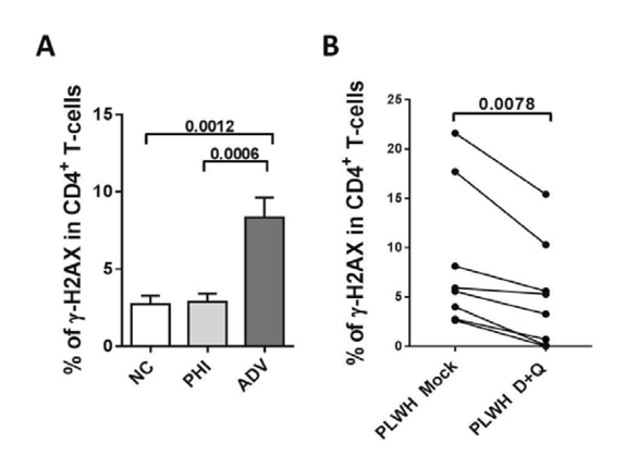
Results: ADV PLWH had a higher amount of Gag+ and CD71+ T-cells, compared with NC (p=0.0006) and ART was able to reduce this activation. SC markers such as SA-βGal, p16INK4a, γ-H2AX (Fig 1A) and Bcl-II were increased in CD4+ (p<0.05) and CD8+ T-cells, especially in ADV vs PHI and NC, and ART do not drop their levels. IL-6 was also higher in CD4+ and CD8+ T-cells from ADV, and in contrast with SC markers, ART normalized these levels (p<0.05). In ADV, a higher proportion of Gag+ and IL-6+ monocytes (M) were observed, whereas CD87+ expression decreased in M. IL-6+ M directly correlate with CD4+ T cell expressing SC biomarkers such as γ-H2AX (p=0.023), p16INK4a (p=0.012) and Bcl-II (p=0.031), but inversely correlated with CD87+-M (r=-0.566 p=0.0004). D+Q senolytic drugs specifically reduced the expression of SC markers as SA-βGal, γ-H2AX (Fig 1B, p=0.0078) and IL-6 in CD4+ T cells. This fall was coupled to a rise in cell mortality induced by D+Q.
Conclusion: HIV-1 infection raises SC biomarkers in T-cells and increases the amount of IL-6+ monocytes, but reduces CD87 expression in these cells.
ART cannot reverse markers of cellular aging excepting IL-6 levels in T cells.
Ex vivo D+Q senolytic treatment decreased the levels of SC biomarkers suggesting that these drugs could be useful to reverse cellular senescence in PLWH.
|
| |
|
|
|
|
|