| |
Diabetes & HIV - Glucose Intolerance in Persons with HIV is Characterized by Subcutaneous Adipose Tissue Enriched For Lipid-processing Macrophages And Effector Memory T Cells With Discrete Transcriptional Phenotypes (Complications Workshop)
|
| |
| |
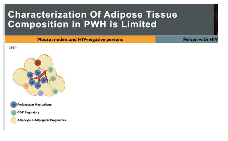
Adipose tissue has a major role in energy intake, energy storage, and lipid and glucose metabolism. Much of what we know about adipose tissue and in particular, the immune cells present in adipose tissue comes from mouse models and from HIV-negative persons. Additionally, most studies to date have used lean and obese comparisons to infer the changes that occur with metabolic disease. This graphic has been simplified for this. In lean individuals, perivascular or alternatively activated macrophages predominate and are generally considered anti-inflammatory. Additionally, CD4 T regulatory cells are abundant. Adipocytes are smaller and progenitor cells have adipogenic capacity. With obesity, CD8 T cells infiltrate adipose tissue, and macrophages polarize towards lipid-processing phenotype. Additionally, CD4 Th1 cells increase. Adipocyte necrosis occurs with hypoxia, which release inflammatory lipids. Overall there is a reduction in the anti-inflammatory cell populations seen in lean adipose tissue. Less is known about adipose tissue in persons with HIV. In PWH without obesity, CD8 T cells infiltrate adipose tissue, there is increased injury and fibrosis of the AT, and immune cells that serve as latent viral reservoirs may contribute to inflammation. In PWH with glucose intolerance, cytotoxic CD4 T cells expand and macrophage inflammatory cytokines increase. However, a global understanding of the cell types that compose AT in PWH to date has been missing.
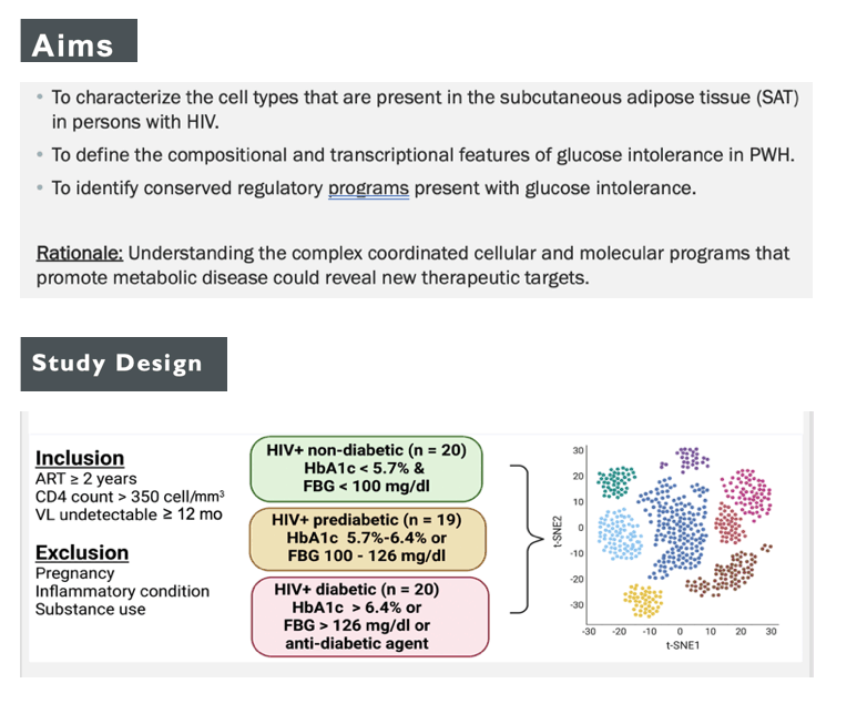
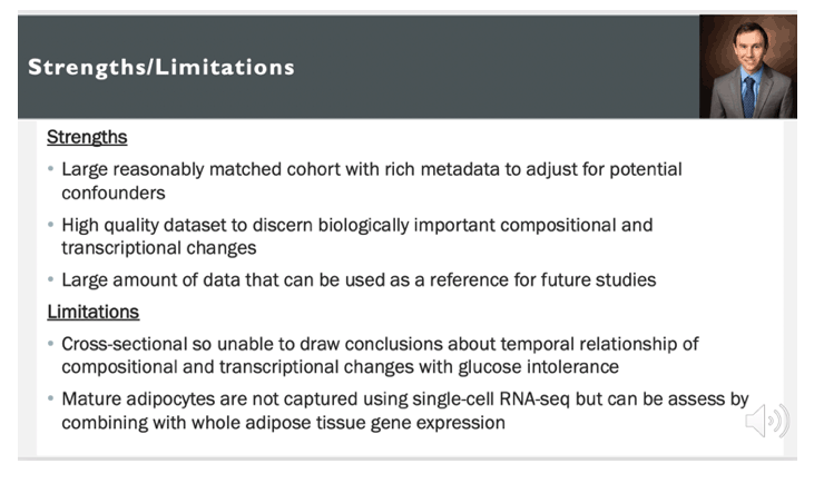
The participants were a subset from a cohort, established to understand the immune factors that are associated with metabolic disease in PWH. Individuals included in the study were on antiretroviral therapy for at least 2 years, had a CD4 count > 350 cells/mm3 at enrollment, and had a viral load that was undetectable for at least 12 months. Individuals were excluded if they were pregnant, had an inflammatory condition, or had self-reported substance use. Participants were categorized by metabolic status based on hemoglobin A1c, fasting blood glucose, and use of anti-diabetic agent. A total of 59 participants underwent subcutaneous adipose tissue biopsy for single-cell RNA sequencing analysis.
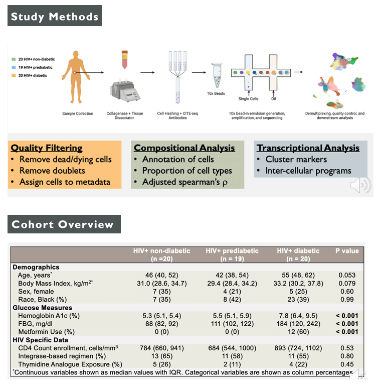
Overall, we included 20 PWH without diabetes, 19 PWH with prediabetes, and 20 PWH with diabetes. PWH with diabetes trended towards older age and high BMI, but no difference in sex or race. As expected, measures of fasting blood glucose, hemoglobin A1c, and metformin use were significantly higher in PWH with diabetes. The majority of participants were on an INSTI-based regimen and a small proportion had exposure to older thymidine analogues.
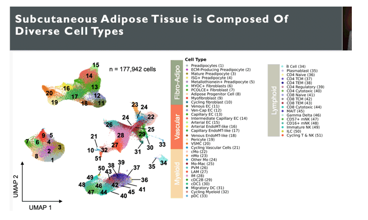
The first aim was to identify the cell types present in SAT in PWH. After a significant amount of quality control filtering, our dataset included 177,942 cells from 59 PWH. We identified 55 distinct cell types, some of which were collapsed in this figure for visualization. Here we can broadly see clustering of cells by type: fibro-adipogenic, vascular, myeloid, B cells, and T cells and NK cells. Our unbiased single-cell RNA and CITE-seq captured a diversity of cells, which reflects the diverse and broad function of adipose tissue.
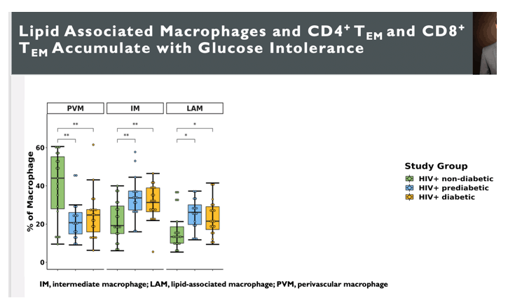
We first examined differences in composition by metabolic status, as this likely reflects functional changes in the adipose tissue. In agreement with mouse models and studies of HIV-negative persons with obesity, the proportion of perivascular macrophages or PVM in those with prediabetes and diabetes was lower than those without diabetes. In contrast, the proportion of lipid-associated macrophages or LAMs and intermediate macrophages or IMs, were increase in those with prediabetes and diabetes compared to those without diabetes. Additionally, the proportions of CD4+ and CD8+ TEM cells was higher in those with prediabetes compared to those without diabetes.
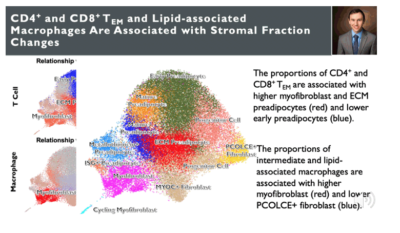
One major question is does the immune cell composition influence the stromal cell composition. Here is the stromal fraction, specifically the fibroblast and adipogenic cells showing populations of fibroblast, myofibroblast, progenitor cells, and preadipocytes. We used spearman correlation between the proportion of CD4 and CD8 TEM shown on the left and right with stromal cell populations. Here we show that the proportion of CD4 and CD8 TEM are associated with higher extracellular matrix producing preadipocytes and lower early preadipocytes. CD4 TEM specifically were also associated with higher myofibroblast. We did the same thing for intermediate macrophages and lipid-associated macropahges, which showed that the proportions of these macrophages were associated with higher myofibroblast and lower PCOLCE+ fibroblast. PCOLCE+ fibroblast are interstitial fibroblast that give rise to preadipocytes. The take away is that immune cells that as associated with glucose intolerance do influence the stromal composition with a shift towards fibrotic cells types.
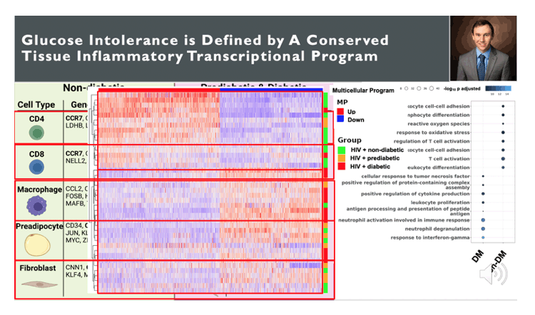
Finally, cell expression changes with glucose intolerance do not occur in isolation among different cell types. Assessing the transcriptional changes across cell types with glucose intolerance could yield insight into tissue level expression changes that occurs with glucose intolerance. To do this, I used dialogue which is a program that finds highly correlated gene expression profiles across cell types that are related to the disease of interest, in this case glucose intolerance. Here is a visual representation with genes that are up in the multicellular program in red and genes that are down in the multicellular program in blue. The columns are gene expression that has been normalized and scaled. The rows are individuals, which are ploted with hierarchical clustering. Higher gene expression of genes that are up in the multicellular program are predominantly non-diabetic where as higher expression of genes that are down in multicellular program are predominantly prediabetic and diabetic. We can look at individual cell types in non-diabetic and prediabetic and diabetic individuals. Among CD4 T cells, non-diabetic individuals had higher expression of CCR7 and SELL, migration genes while diabetic had expression of IFNG, TNF, cytotoxic, and activation genes. CD8 T cells had similar expression profile as CD4 T cells. Macrophages in non-diabetic individuals expressed higher levels of chemokines as well as transcription factors associated with perivascular macrophage polarization. In contrast, pre-diabetic and diabetic individuals had expression of lipid processing genes including TREM2, APOC1, APOE. Preadipocytes in non-diabetic individuals expressed markers of adipogenesis including CEBPB and EGR1 whereas in diabetics, they expressed genes associated with fibrosis in POSTN. This pattern was also seen in ifbroblasts. Finally, over representation analysis of genes associated with diabetic individuals show response to IFNG, neutrophil degranulation, TNFA wherease genes associated with non-diabetic individuals were related to leukocyte regulation and cytokines/chemokines.
|
|
| |
| |
|
|
|