 |
|
|
| |
Infected T-Cell Clones Are Shared Across CSF and Blood Compartments in PWH
|
| |
| |
CRO 2024 March 3-6 Denver
Meng Wang, Jennifer Yoon, Hailey Reisert, Bibhuprasad Das, Jennifer Chiarella, John W. Mellors, Alina P. Pang, Joshua C. Cyktor, Margaret Fikrig, Elias K. Halvas, Yuval Kluger, Serena Spudich, Michael J. Corley, Shelli Farhadian
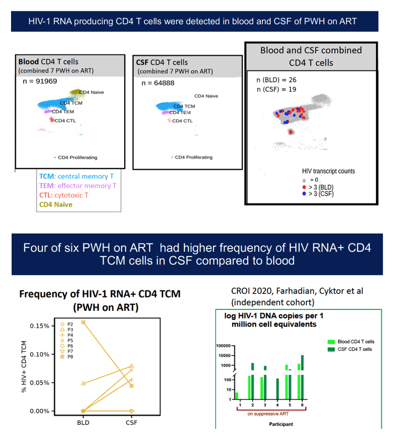
Background: The central nervous system (CNS) is a site of persistently infected cells during HIV infection. However, the dynamics of CNS infection and T cell trafficking in PWH are incompletely understood. Here, we utilized single-cell T cell receptor (TCR) and transcriptome profiling of paired CSF and blood from PWH to gain insights into the dynamics of rare HIV-1 RNA-producing T cells in both compartments, and under the pressure of ART.
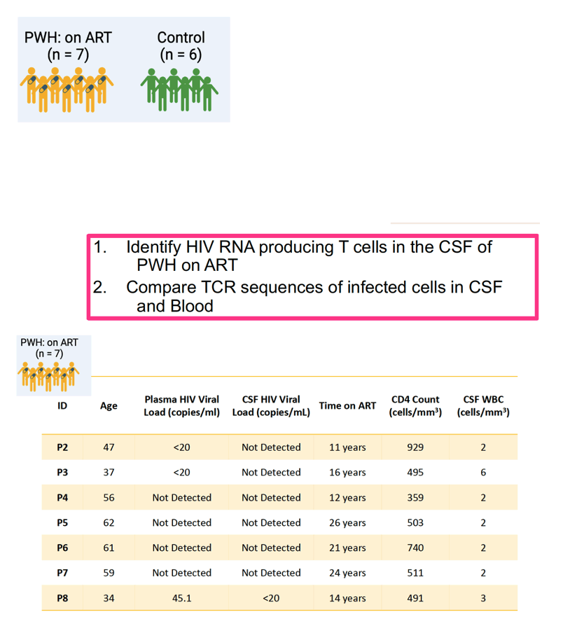
Methods: We enrolled eight PWH; seven were on suppressive ART and one had chronic HIV profiled before and 3, 7, and 9 months after ART. We also enrolled six HIV-uninfected controls, demographically matched to PWH. We profiled single cell TCR and RNA from paired CSF and blood using 5' V(D)J 10x Genomics scRNA- seq and scTCR-seq. To identify whether there were detectable transcriptionally active HIV-1 RNA producing cells in CSF and blood, we aligned the single cell transcriptome sequencing reads against consensus and autologous HIV-1 genomes.
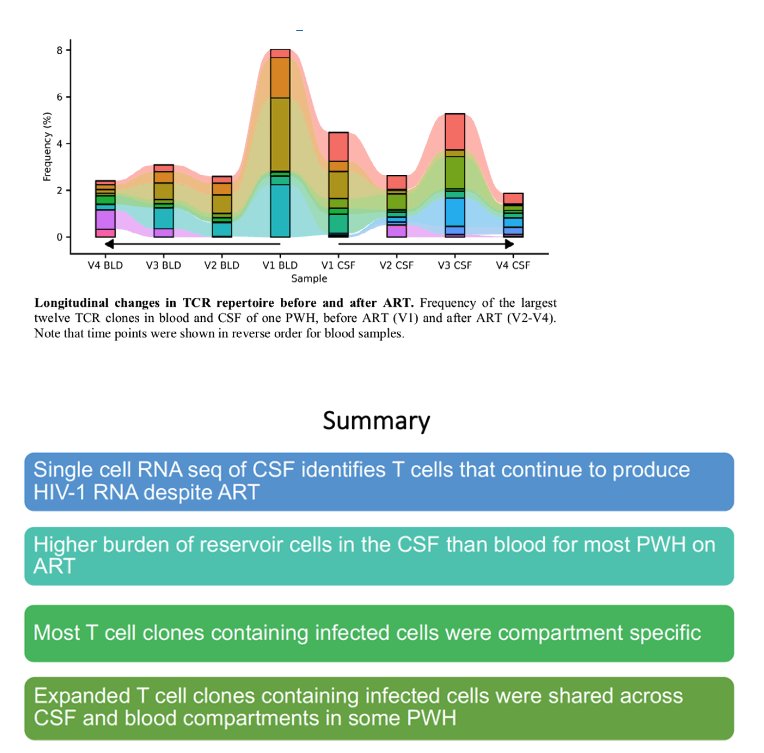
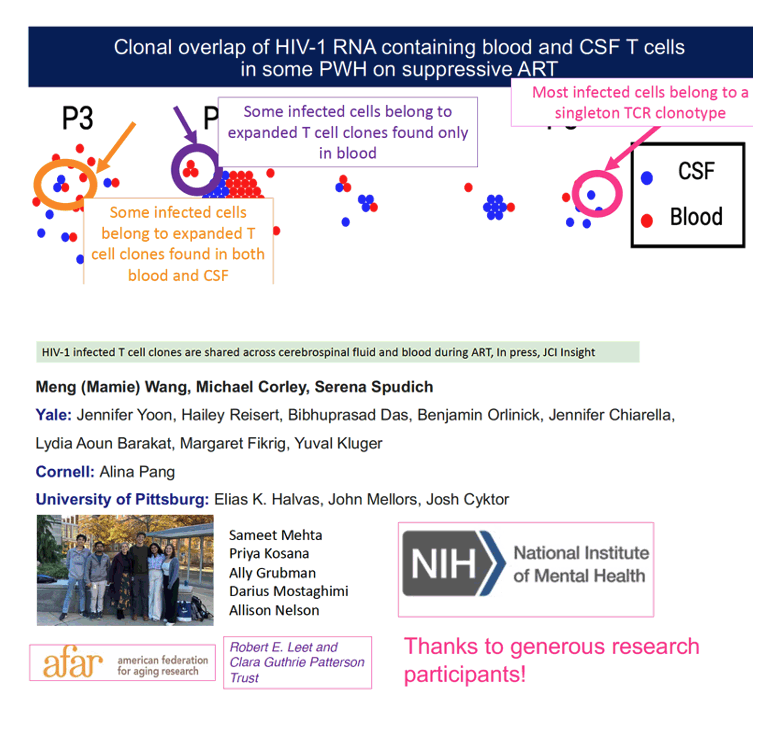
Results: In total, we examined the single-cell transcriptomes of 129,544 CSF cells and 262,818 PBMCs from PWH and controls. We detected transcriptionally active HIV-1 RNA producing cells in 8/11 (72.7 %) CSF samples and 6/11 (54.5 %) blood samples, with a higher frequency of infected single CD4+ T cells in CSF than in blood. Among infected CD4+ T cells, a majority (83.6 %) were identified as CD4+ central memory T cells. Differential expression analyses revealed infected CSF T cells displayed a unique transcriptional profile compared to uninfected CSF T cells. We utilized scTCR data to identify 36 T cell clones containing infected cells. Most (78%) of these T cell clones were tissue specific (found in blood or CSF but not both), but some (22%) clones containing infected cells were found in both CSF and blood. Most infected cells belonged to singletons (unique TCR clones), but 28% belonged to TCR clones with evidence of clonal expansion. Longitudinally following one PWH before and at three time points after initiating ART, we found infected T cell clones that persisted after ART initiation, in both CSF and blood, including a T cell clone that expanded in the CSF several months after ART initiation.
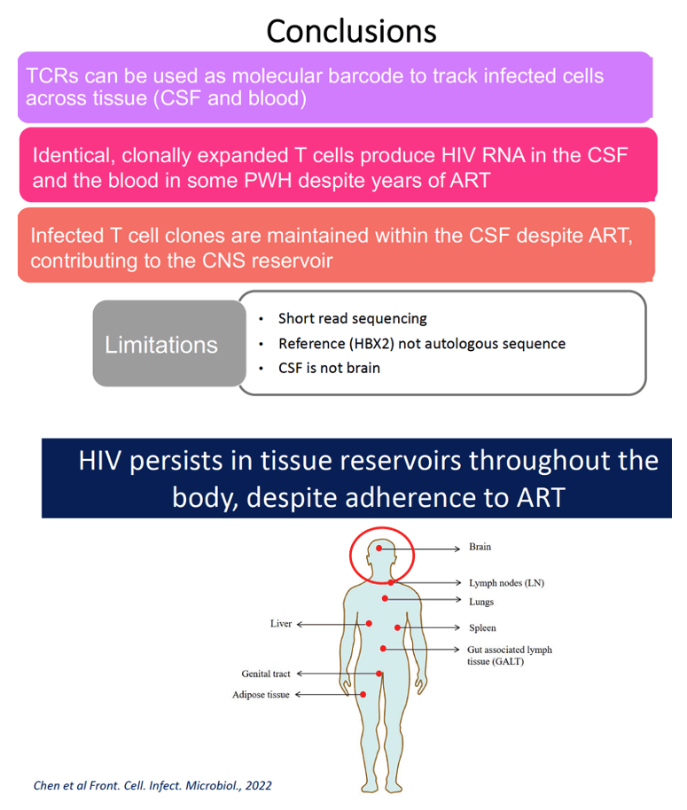
Conclusion: By tracking T cell clones across times and tissue, we find that T cell clones persist in the CNS over time. Infected, identical, and expanded T cell clones are found across tissue compartments. Our findings suggest that maintenance and expansion of infected T cell clones contributes to the CNS reservoir in PWH on ART.
|
| |
|
|
|
|
|